Science

LRP1 is a master regulator of tau uptake and spread

Our latest publication in the journal Nature is now avaliable!

Authors: Jennifer N. Rauch, Gabriel Luna, Elmer Guzman, Morgane Audouard, Collin Challis, Youssef E. Sibih, Carolina Leshuk, Israel Hernandez, Susanne Wegmann, Bradley T. Hyman, Viviana Gradniaru, Martin Kampmann, and Kenneth S. Kosik

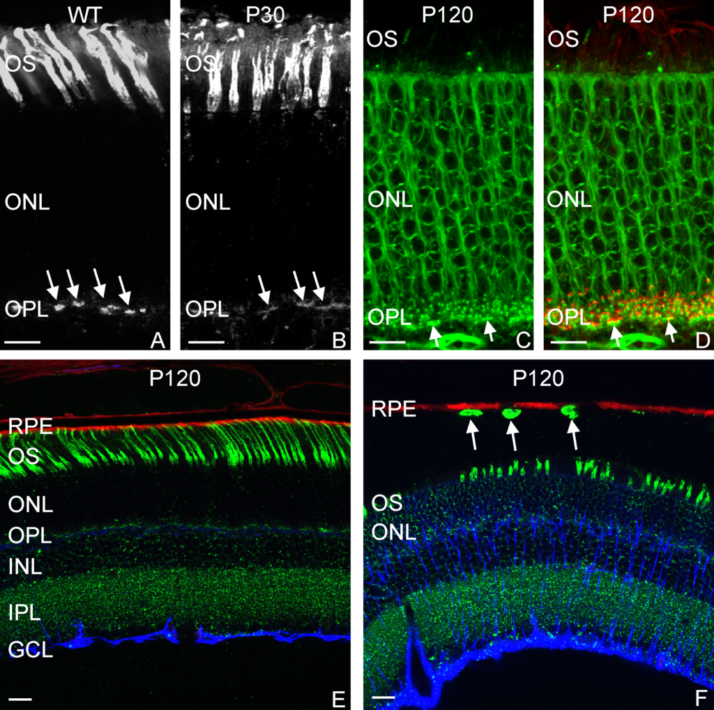
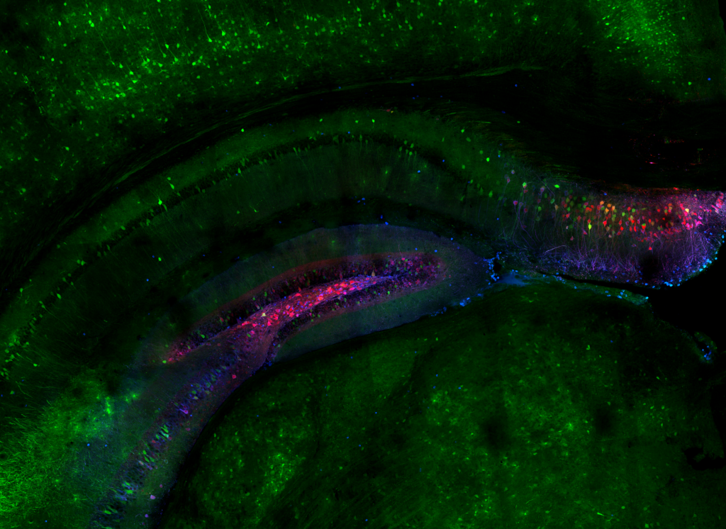
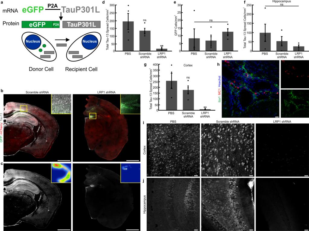
Abstract:  The spread of protein aggregates during disease progression is a common theme underlying many neurodegenerative diseases. The microtubule-associated protein tau has a central role in the pathogenesis of several forms of dementia known as tauopathies—including Alzheimer’s disease, frontotemporal dementia and chronic traumatic encephalopathy. Progression of these diseases is characterized by the sequential spread and deposition of protein aggregates in a predictable pattern that correlates with clinical severity. This observation and complementary experimental studies have suggested that tau can spread in a prion-like manner, by passing to naive cells in which it templates misfolding and aggregation. However, although the propagation of tau has been extensively studied, the underlying cellular mechanisms remain poorly understood. Here we show that the low-density lipoprotein receptor-related protein 1 (LRP1) controls the endocytosis of tau and its subsequent spread. Knockdown of LRP1 significantly reduced tau uptake in H4 neuroglioma cells and in induced pluripotent stem cell-derived neurons. The interaction between tau and LRP1 is mediated by lysine residues in the microtubule-binding repeat region of tau. Furthermore, downregulation of LRP1 in an in vivo mouse model of tau spread was found to effectively reduce the propagation of tau between neurons. Our results identify LRP1 as a key regulator of tau spread in the brain, and therefore a potential target for the treatment of diseases that involve tau spread and aggregation.

Farnesyl Transferase Inhibition for the Treatment of Tauopathies

Our latest publication in the journal Science Translational Medicine is now avaliable! The article in its entirety can be found here. I was fortunate enough to provide the imaging data for this project as part of the Kosik Molecular and Cellular Neurobiology lab. I was also lucky enough to have my image selected as a the journal cover.

Journal cover Science Translational Medicine, March issue 2019.

Authors: Israel Hernandez, myself, Michel Giroux, Celeste Karch, Daniel Boctor, Youssef Sibih, Nadia Storm, Antonio Dias, Cezary Zekanowski, Alex Kang, Cassidy Hinman, Vesna Cerovac, Elmer Guzman, Honjun Zhou, Alison Goate, Steve Fisher, Ana Cuervo, Ken Kosik.

Lateral region of the hippocampus showing pyramidal cells in yellow and protoplasmic astrocytes in magenta.

Abstract:
Tau inclusions are a shared feature of many neurodegenerative diseases, among them frontotemporal dementia caused by tau mutations. Treatment approaches for these conditions include targeting posttranslational modifi-cations of tau proteins, maintaining a steady-state amount of tau, and preventing its tendency to aggregate. We discovered a new regulatory pathway for tau degradation that operates through the farnesylated protein, Rhes, a GTPase in the Ras family. Here, we show that treatment with the farnesyltransferase inhibitor lonafarnib reduced Rhes and decreased brain atrophy, tau inclusions, tau sumoylation, and tau ubiquitination in the rTg4510 mouse model of tauopathy. In addition, lonafarnib treatment attenuated behavioral abnormalities in rTg4510 mice and reduced microgliosis in mouse brain. Direct reduction of Rhes in the rTg4510 mouse by siRNA reproduced the results observed with lonafarnib treatment. The mechanism of lonafarnib action mediated by Rhes to reduce tau pathology was shown to operate through activation of lysosomes. We finally showed in mouse brain and in hu-man induced pluripotent stem cell–derived neurons a normal developmental increase in Rhes that was initially suppressed by tau mutations. The known safety of lonafarnib revealed in human clinical trials for cancer suggests that this drug could be repurposed for treating tauopathies.

Selected region of the hippocampus showing resident microglia in green, astrocytes in red, the mutant form of tau in white.
Selected region of the hippocampus showing resident microglia in green, astrocytes in red, the mutant form of tau in blue.

News and Media Coverage: Scientific American, The Conversation, Gizmodo, UC Santa Barbara, San Francisco Chronicle.

2018 Nikon Small World

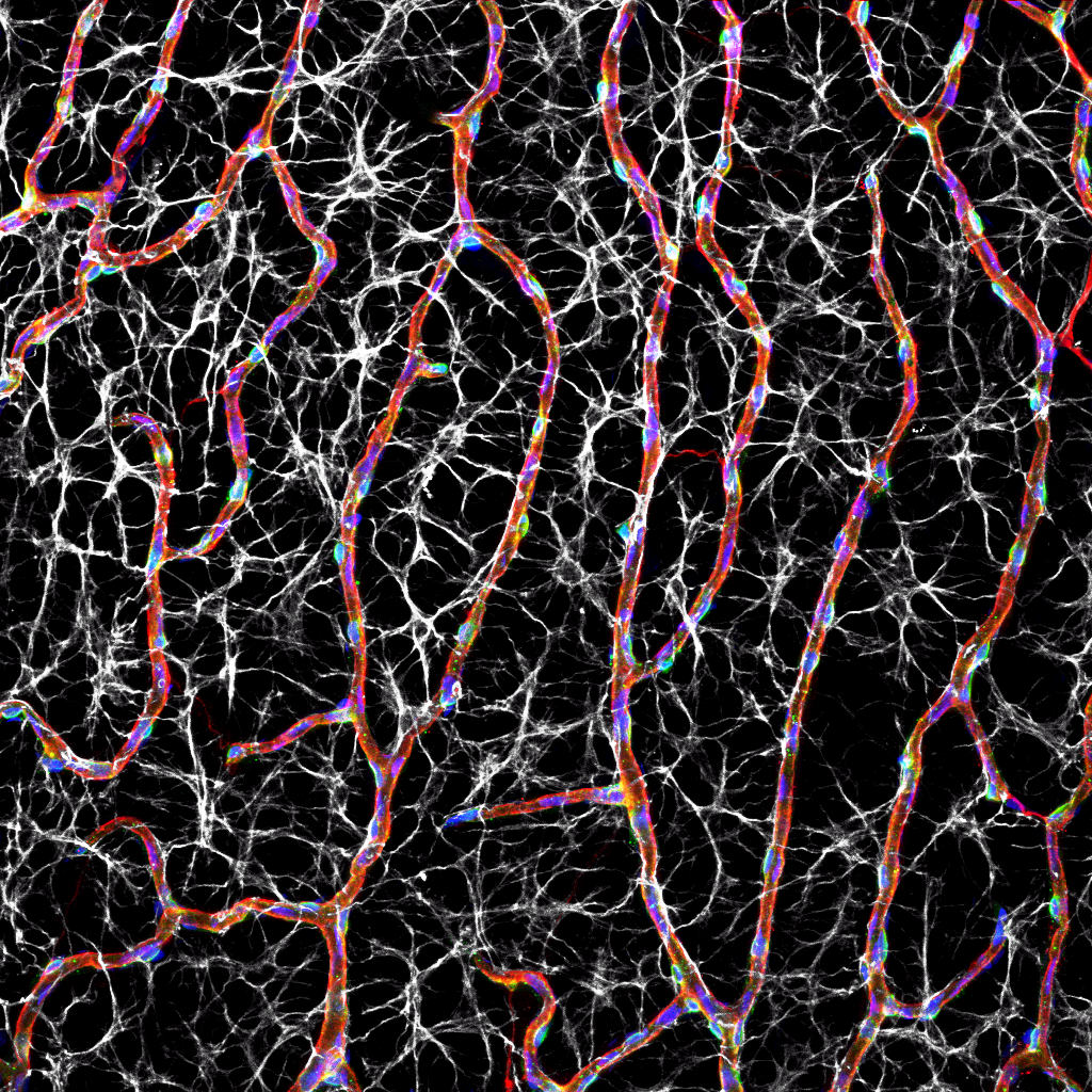
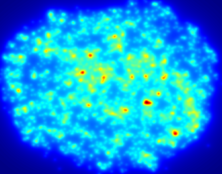
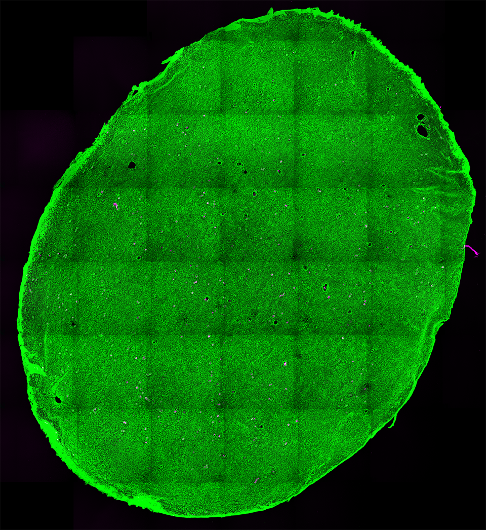
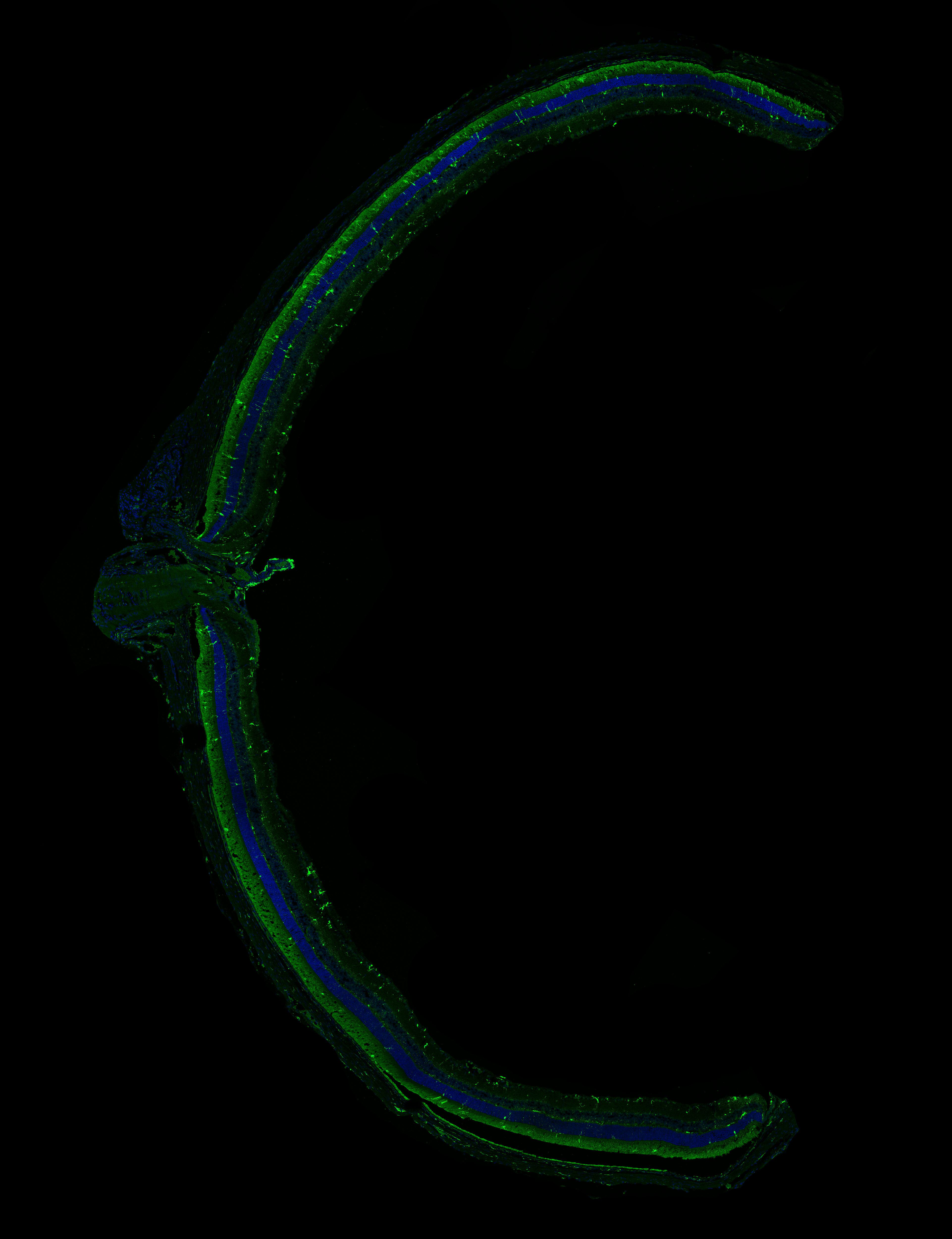
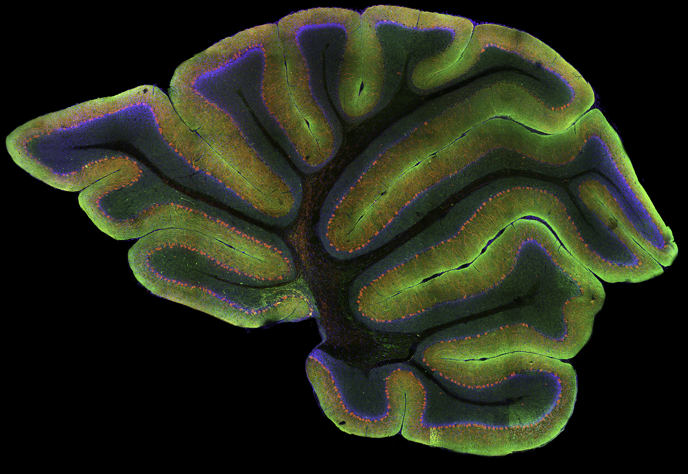
High-resolution, wide-field mosaic of the cerebellum.

Each year I look forward to the Nikon Small World International Photomicrography competition.  This year I was fortunate enough to receive image of distinction honors.  The Small World competition featured thousands of entries from 89 countries, showcasing some of the worlds best microscopists who share their talents in the premiere imaging contest.  I entered the image above of the cerebellum as part of my research in Dr. Kenneth Kosik’s cellular and molecular neurobiology lab in the Neuroscience Research Institute at UC Santa Barbara that was captured using immunoctyochemisty and a laser scanning confocal microscope.  The resultant image is comprised of hundreds of individual images or tiles that are seamlessly aligned and registered to provide both a global overview of the specimen as well as the resolution necessary to interrogate selected regions at the single-cell level.  The mosaic shows the Purkinje cells (red) lining the cerebellum, as well as the ubiquitous protein, tau (green) and nuclei (blue).  I have always been drawn to the cerebellum in part because of its sulci and gyri that demonstrate a remarkable sense of evolutionary complexity and aesthetic beauty.

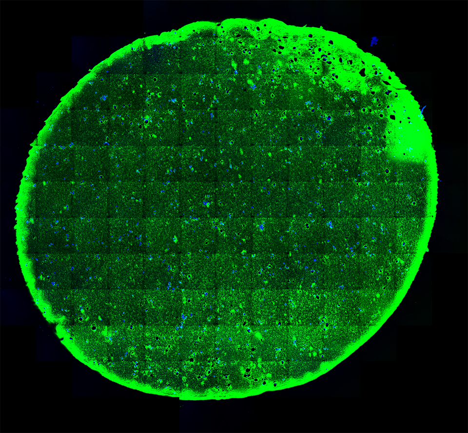
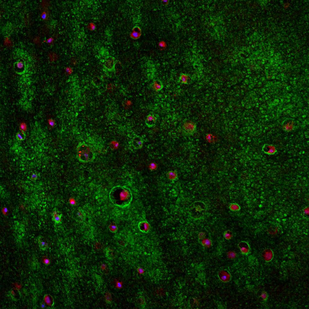
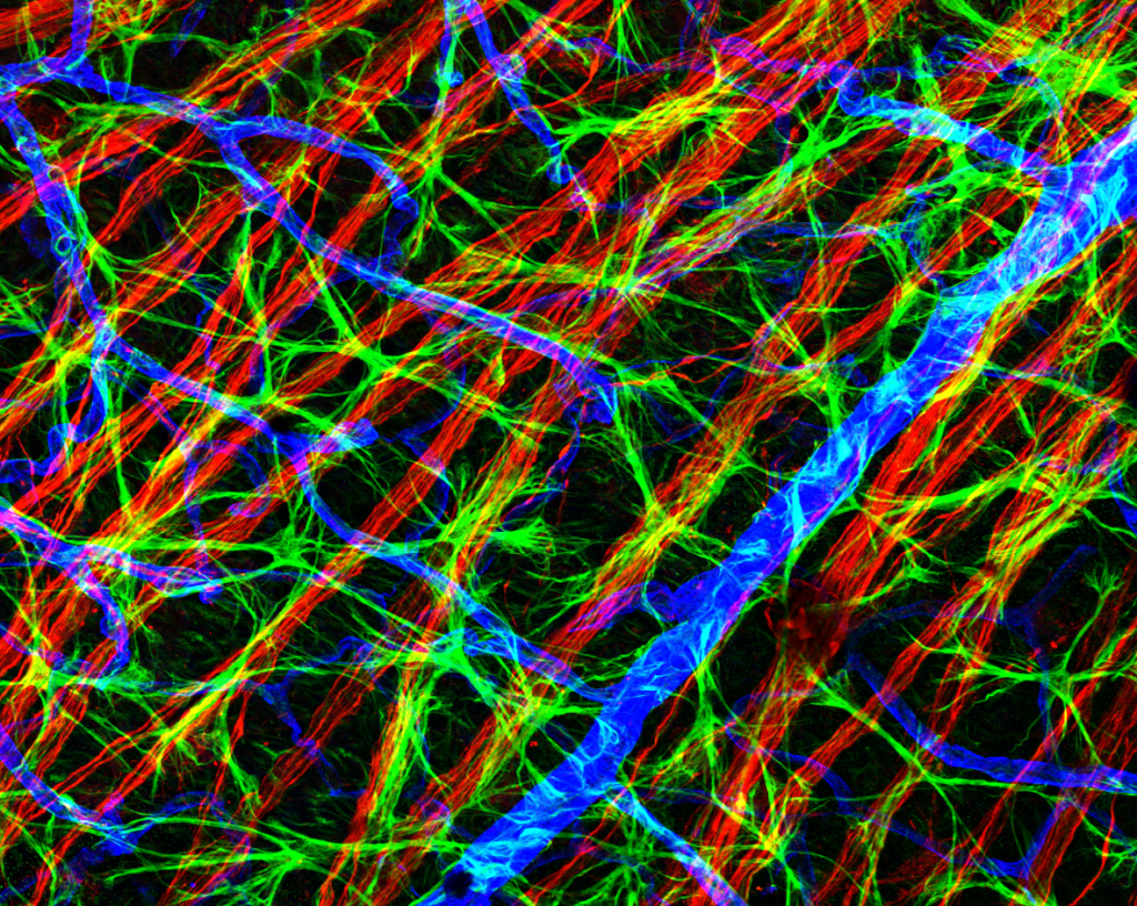
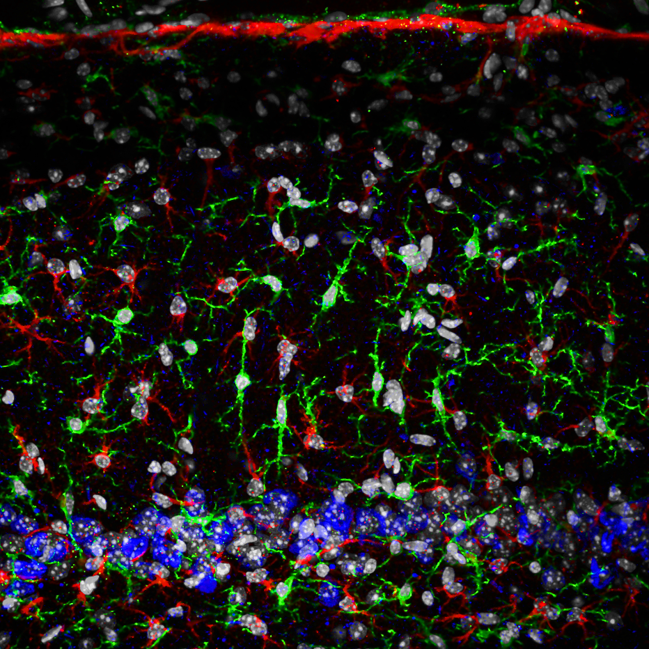
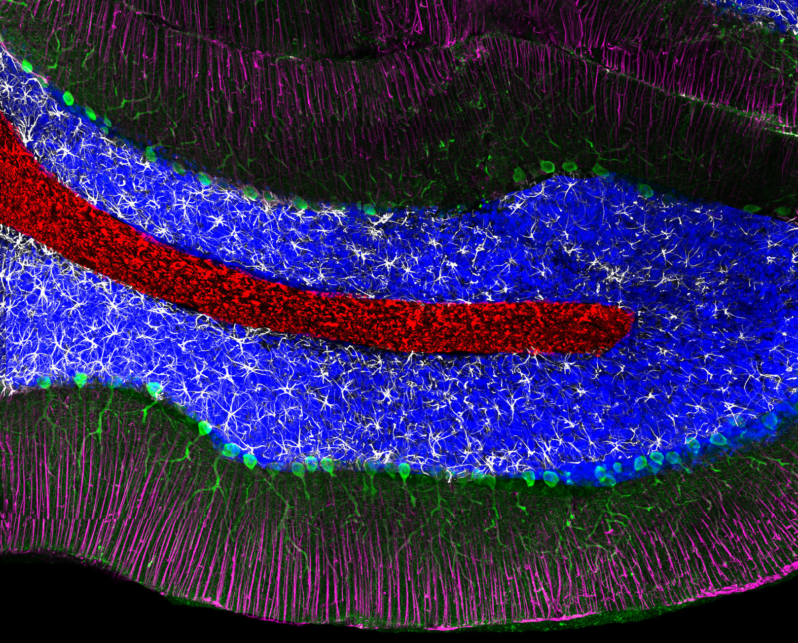
Example of a single tile that comprise the above mosaic.

This example shows the resolution of a single tile that comprises the winning image.  Here, Perkinje cells stained with an antibody to inositol triphosphate 3 (green), the intermediate filament protein GFAP (glial fibrillary acidic protein; white), Bergmann’s glia (magenta), nuclei (blue) and myelin basic protein (red).

Assessment of Outer Retinal Remodeling in the Hibernating 13-lined Ground Squrriel

We have a new publication out in the journal of Investigative Ophthalmology and Visual Science that examined cellular and sub-cellular remodeling in the hibernating ground squirrel.  This research was a result of another fruitful collaboration with Ben Sajdak, a graduate student in the department of advanced ocular imaging program at the medical college of Wisconsin Eye Institute.  Open access PDF can be found here.

Authors: Benjamin S. Sajdak, Brent A. Bell, Taylor R. Lewis, myself, Grayson S. Cornwell, Steven K. Fisher, Dana K. Merriman, and Joesph Carroll.

Abstract:

Purpose. We examined outer retinal remodeling of the euthermic and torpid cone-dominant 13-lined ground squirrel (13-LGS) retina using optical coherence tomography (OCT) imaging and histology.

Methods. Retinas and corneas of living 13-LGSs were imaged during euthermic and torpid physiological states using OCT. Retinal layer thickness was measured at the visual streak from registered and averaged vertical B-scans. Following OCT, some retinas were collected immediately for postmortem histologic comparison using light microscopy, immunofluorescence, or transmission electron microscopy.

Results. Compared to OCT images from euthermic retinae, OCT images of torpid retinae revealed significantly thicker inner and outer nuclear layers, as well as increases in the distances between outer retinal reflectivity bands 1 and 2, and bands 3 and 4. A significant decrease in the distance between bands 2 and 3 also was seen, alongside significant thinning of the choriocapillaris and choroid. OCT image quality was reduced in torpid eyes, partly due to significant thickening of the corneal stroma during this state.

Conclusions. The torpid retina of the hibernating 13-LGS undergoes structural changes that can be detected by OCT imaging. Comparisons between in vivo OCT and ex vivo histomorphometry may offer insight to the origin of hyperreflective OCT bands within the outer retina of the cone-dominant 13-LGS.

Olympus flouview 1000 laser scanning confocal microscope.

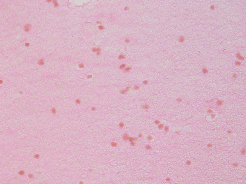
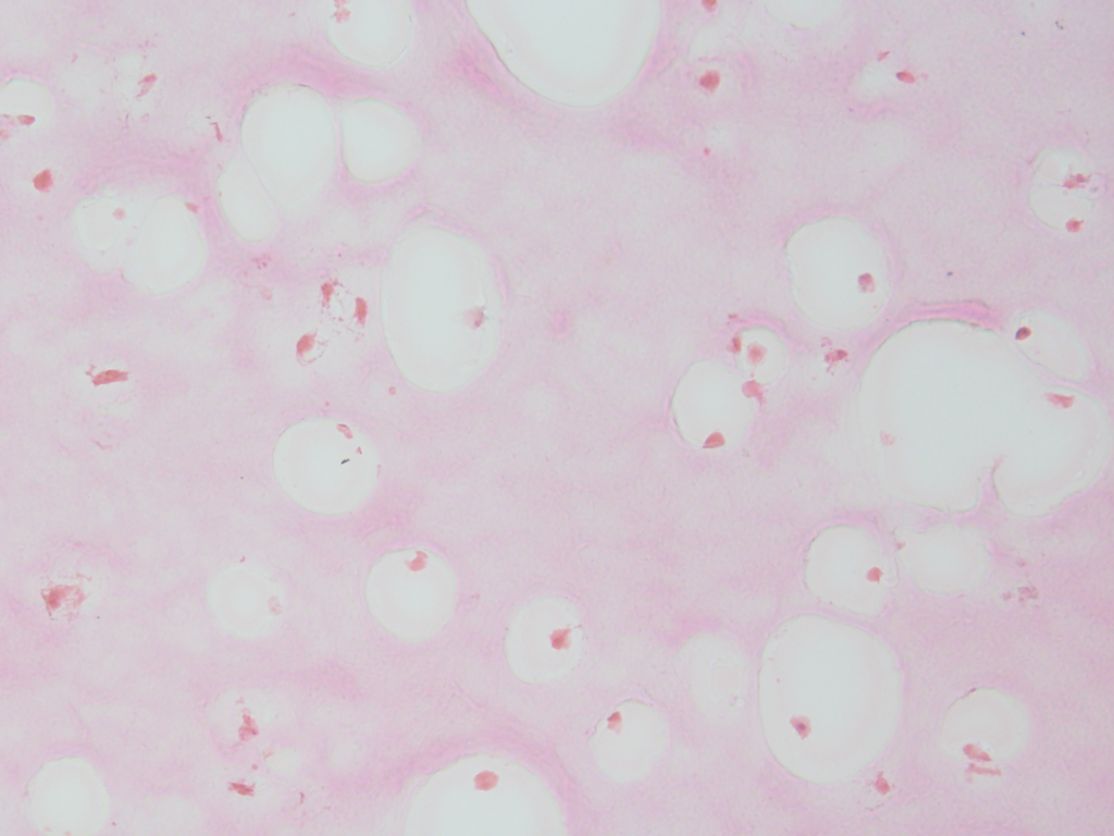
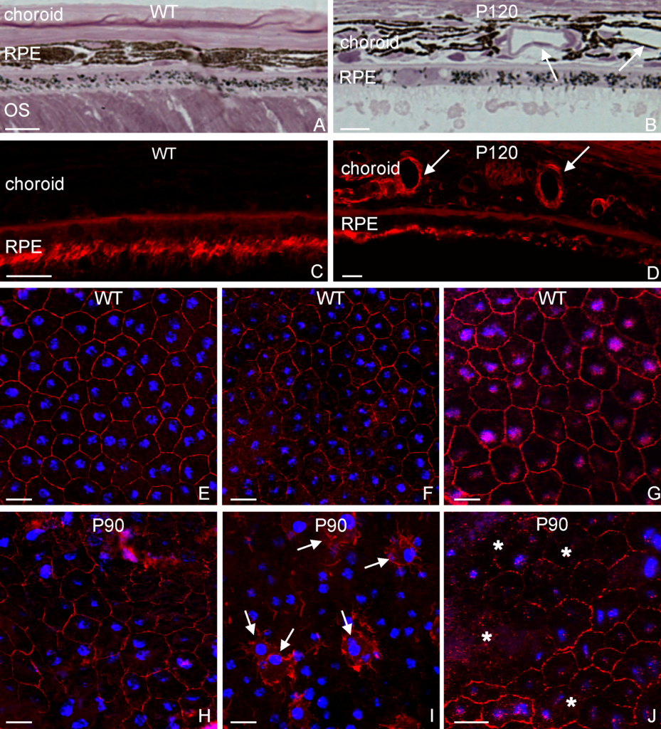
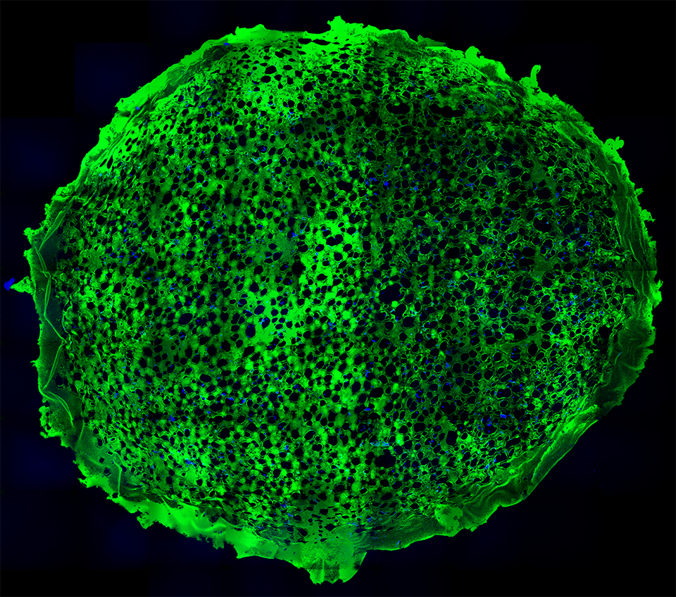
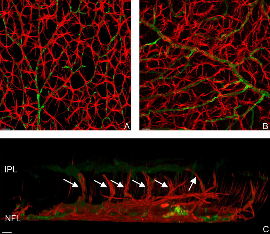
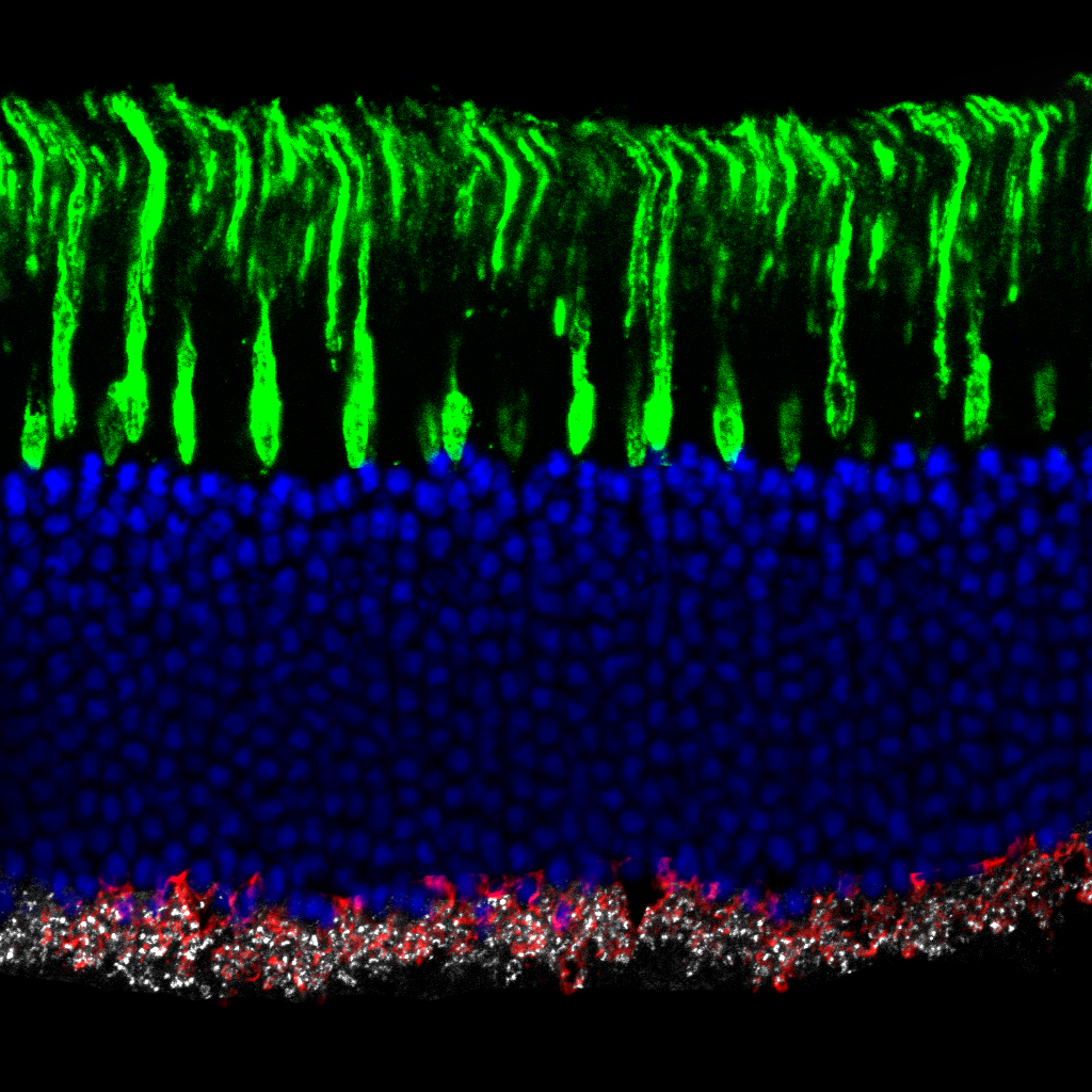
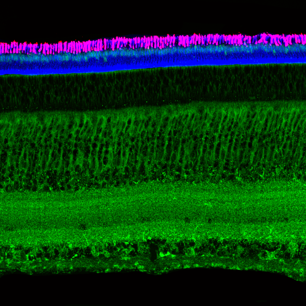
Light micrograph using immunocytochemistry to visualize the cone photoreceptors of the neural retina, magnified 400 times.

Electron micrograph of the light sensitive photoreceptors, magnified 10,000 times.

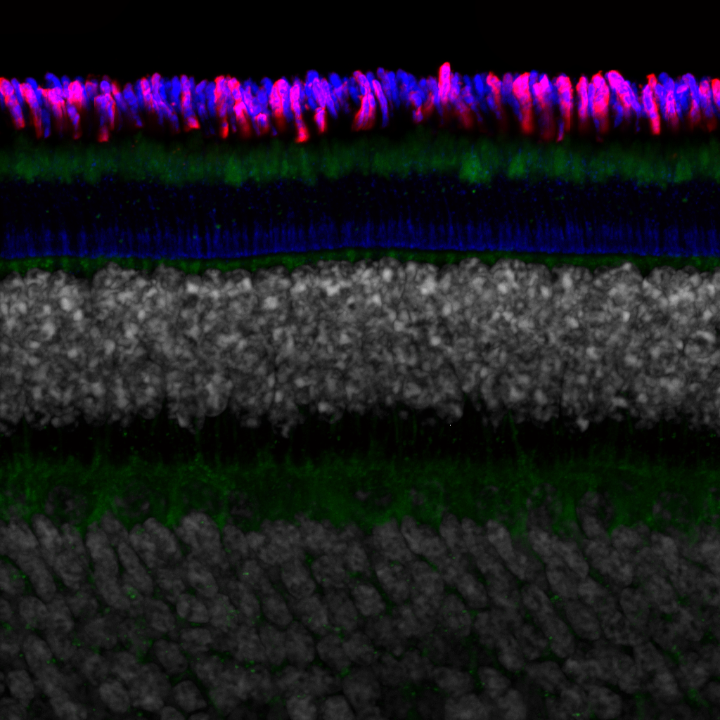
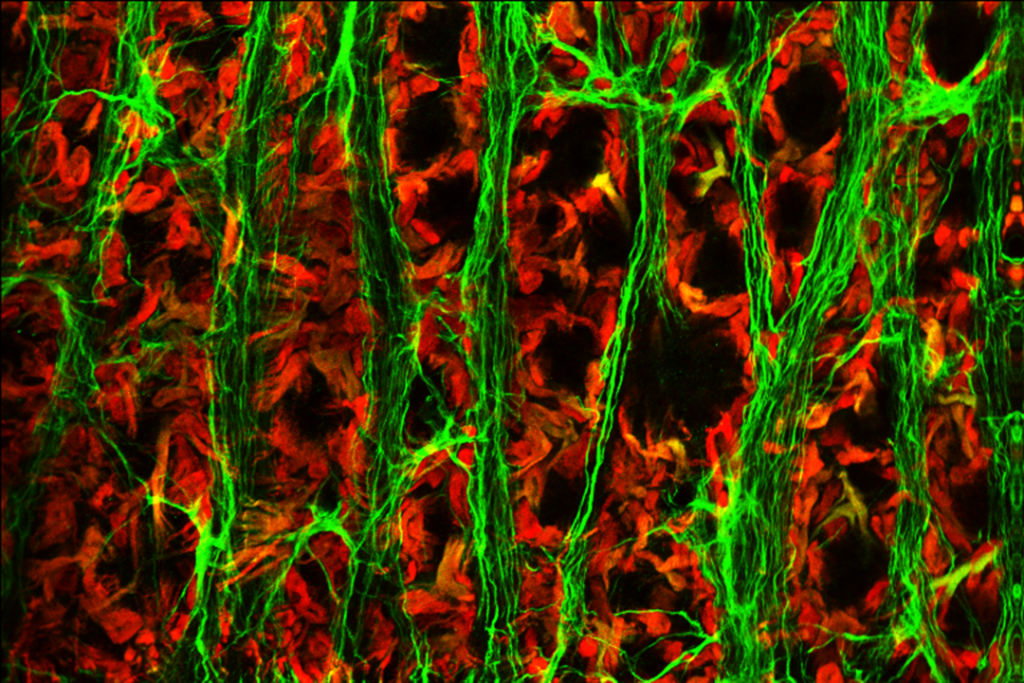
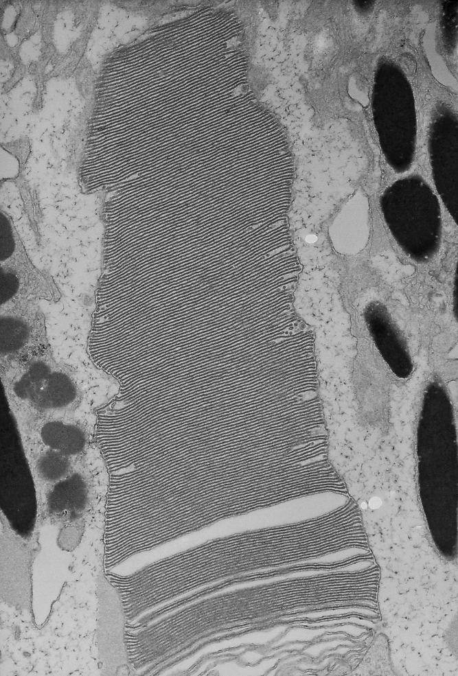
Electron micrograph of a photoreceptor outer segment, magnified 20,000 times.
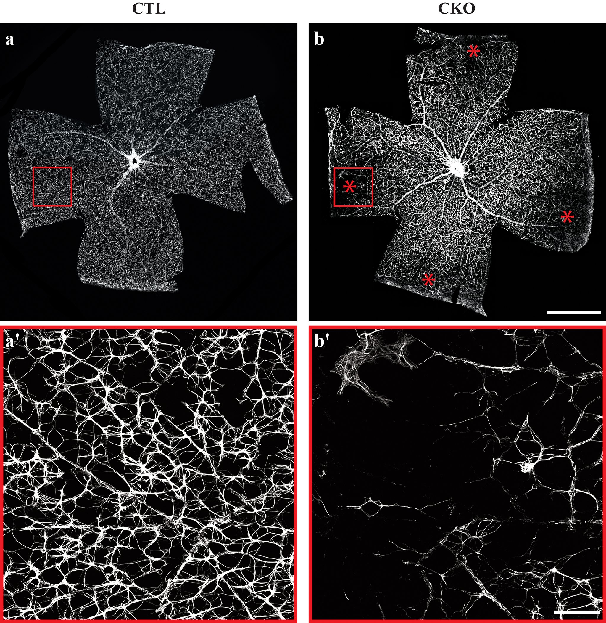
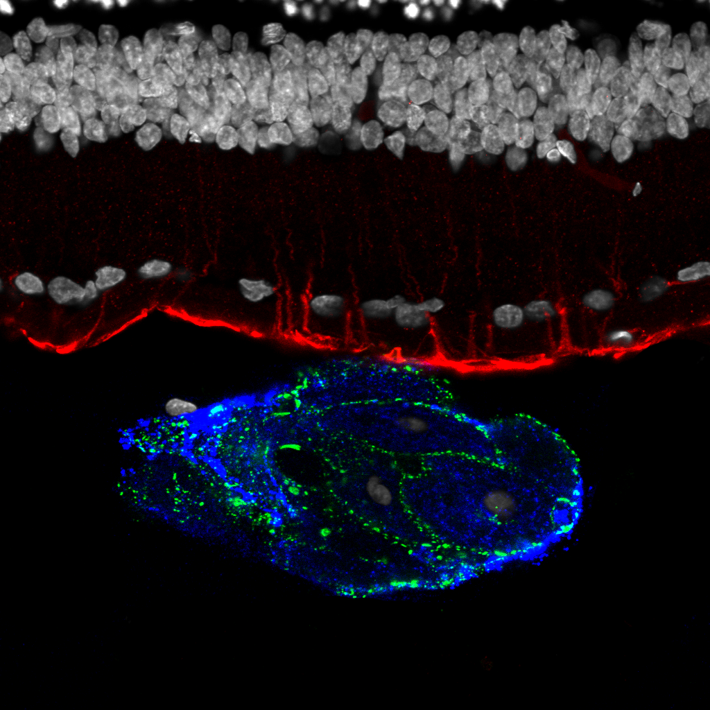
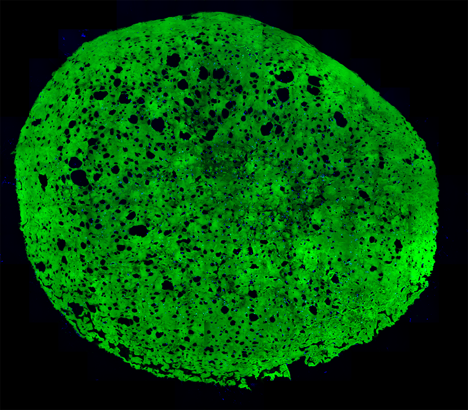
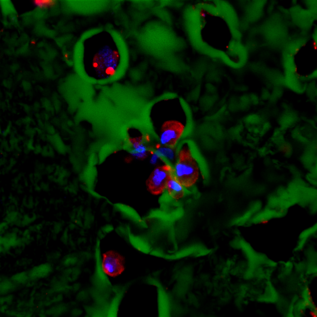
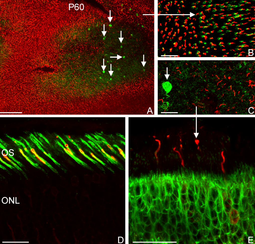
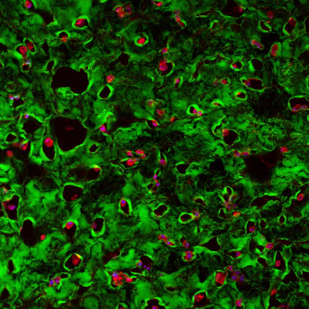
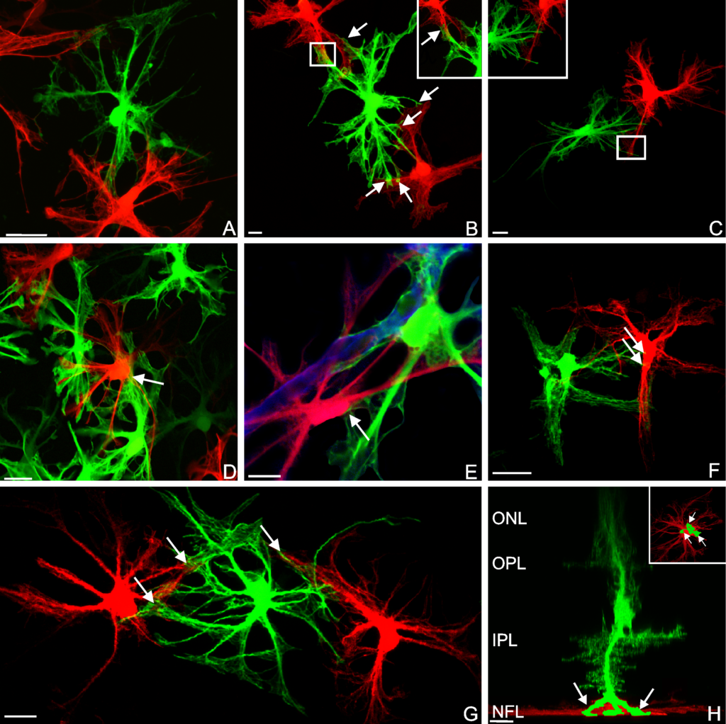
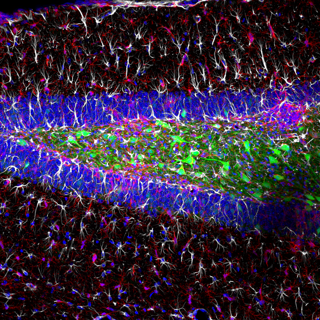
Sox2 Regulates Astrocytic and Vascular Development in the Retina

We have a new publication out in the journal glia that examines the role of the gene Sox2 on the development of astrocytes and the vasculature in the retina.  Read the full article here.

Authors: Amanda Kautzman, Patrick Keeley, Michael Nahmou, myself, Steven Fisher, Benjamin Reese

Abstract:  Sox2 is a transcriptional regulator that is highly expressed in retinal astrocytes, yet its function in these cells has not previously been examined. To understand its role, we conditionally deleted Sox2 from the population of astrocytes and examined the consequences on retinal development. We found that Sox2 deletion does not alter the migration of astrocytes, but it impairs their maturation, evidenced by the delayed upregulation of glial fibrillary acidic protein (GFAP) across the retina. The centro-peripheral gradient of angiogenesis is also delayed in Sox2-CKO retinas. In the mature retina, we observed lasting abnormalities in the astrocytic population evidenced by the sporadic loss of GFAP immunoreactivity in the peripheral retina as well as by the aberrant extension of processes into the inner retina. Blood vessels in the adult retina are also under-developed and show a decrease in the frequency of branch points and in total vessel length. The developmental relationship between maturing astrocytes and angiogenesis suggests a causal relationship between the astrocytic loss of Sox2 and the vascular architecture in maturity. We suggest that the delay in astrocytic maturation and vascular invasion may render the retina hypoxic, thereby causing the abnormalities we observe in adulthood. These studies uncover a novel role for Sox2 in the development of retinal astrocytes and indicate that its removal can lead to lasting changes to retinal homeostasis.

Le retine

The retina is the light-sensitive layer of the eye, responsible for initiating the cascade of events that ultimately leads to visual perception. Vision relies on two types of photoreceptors; rods, used in low-light conditions, and cones (green colored structures in the image above) used for high acuity vision as well as the detection of different wavelengths of radiation (color).  Both are highly specialized cell types that posses the ability to convert photons of light into electrochemical signals that are relayed to the lateral geniculate nucleus and then onto higher centers in the visual cortex where humans experience visual perception.   Under certain conditions (i.e., inherited genetics or physical trauma) photoreceptor degeneration can lead to a gradual decline in visual acuity, and in extreme cases blindness.

To treat certain types sight-threatening conditions, we have begun testing the efficacy of intraocular injections of stem cells (blue and green ovoid structure in the image below) as a means of ameliorating the effects of photoreceptor degeneration, thus preserving vision.

As part of this particular project, I investigated whether intraocular injections of stem cells elicited a response from the immune cells in the retina.  Here, I used a laser scanning confocal microscope to capture many high-resolution images to generate a composite image the entire neural retina (image below).

Composite image.

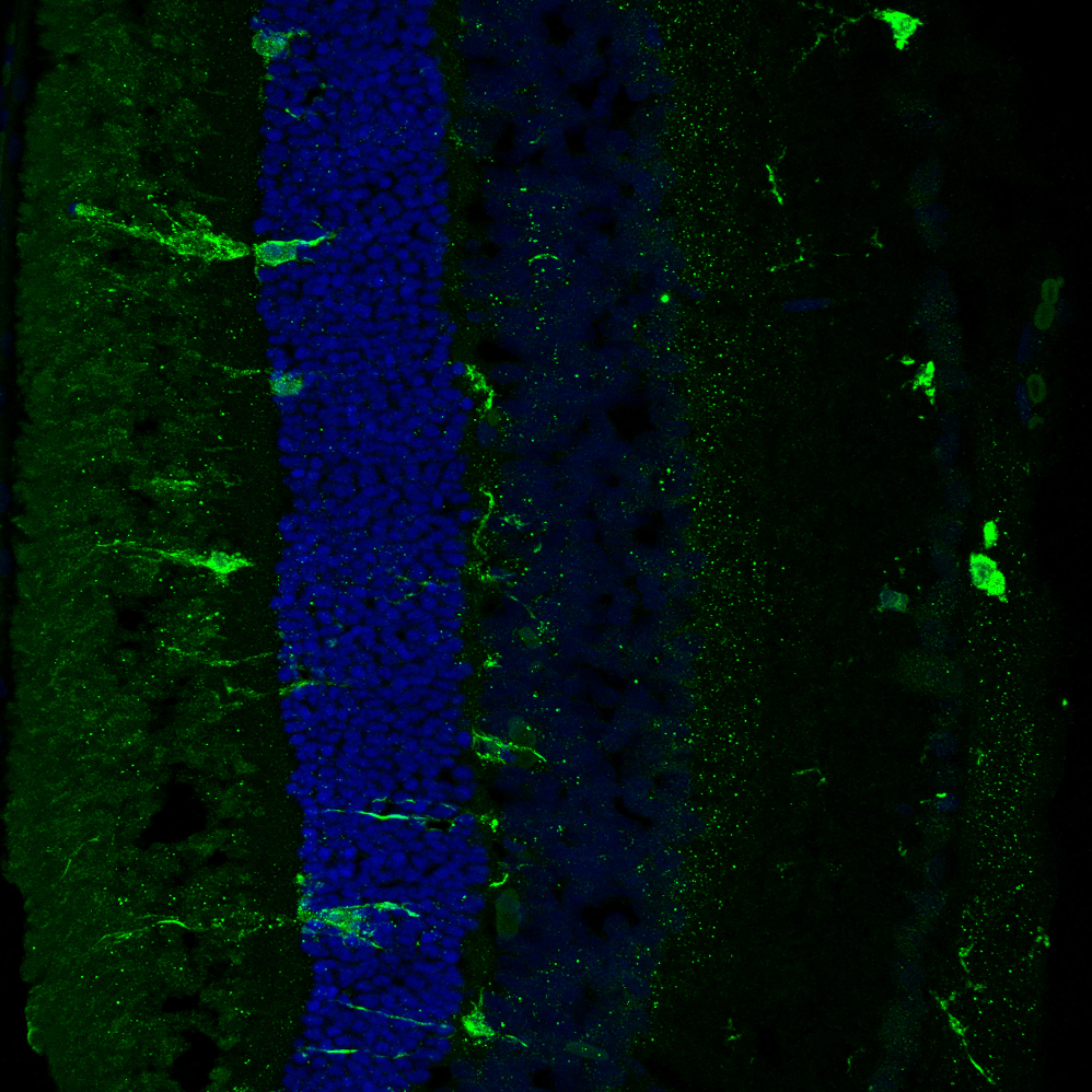
Magnified 400 times.

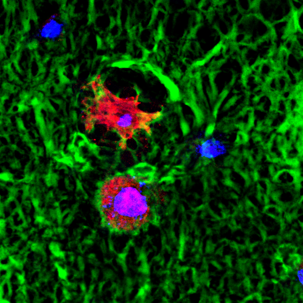
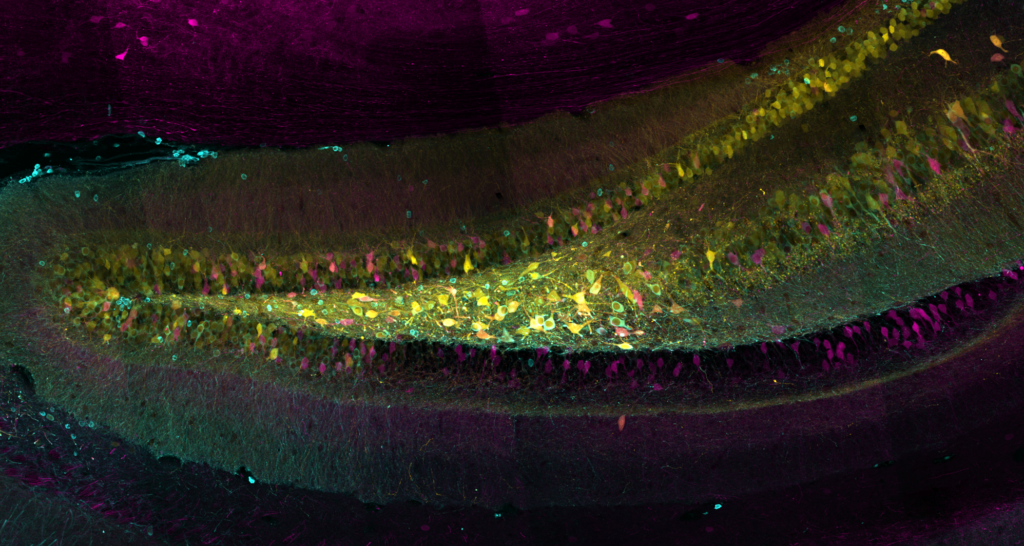
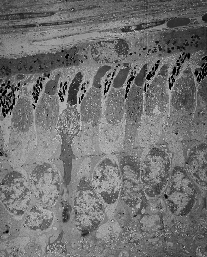
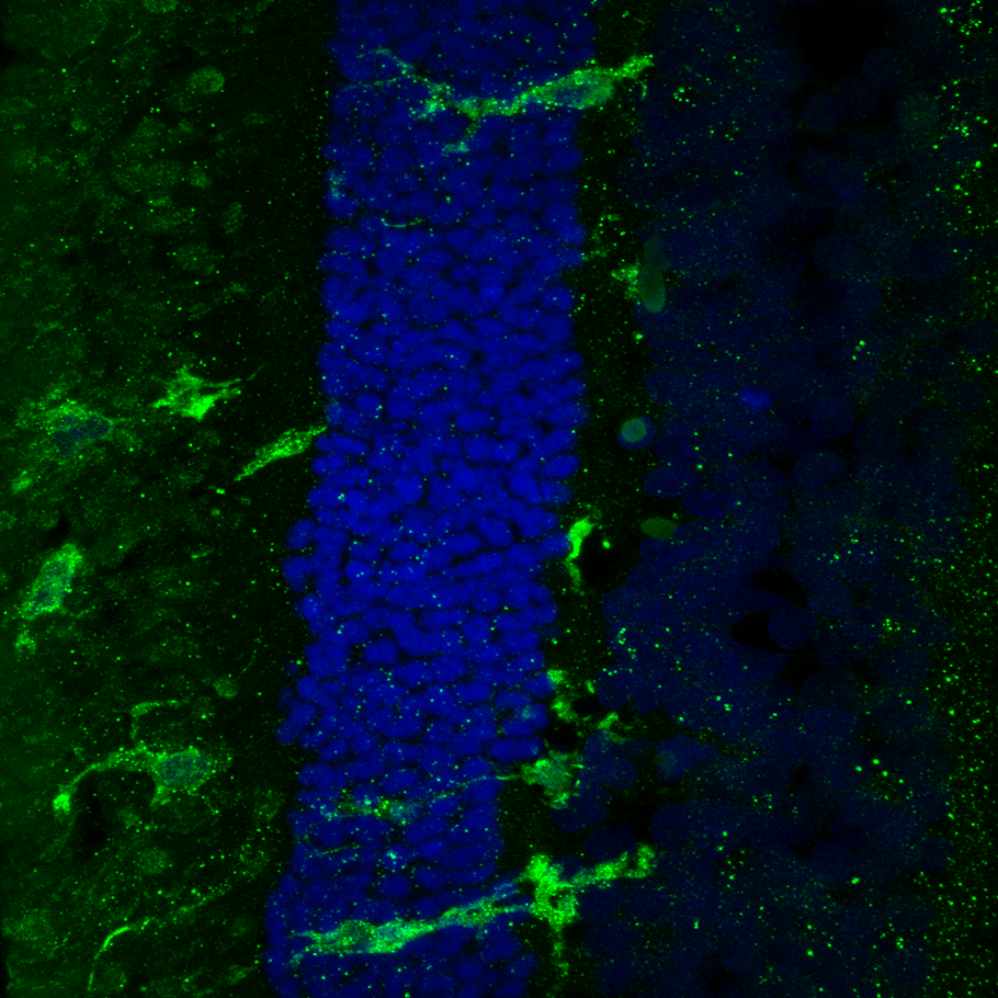
Magnified 600 times.

In these sample images the larger green ‘blobs’ reveal immune cells as they infiltrate the retina in response to neural degeneration.   The blue color in these images shows the layer of photoreceptor nuclei.  While much remains to be seen about whether these cell-based therapies have a positive impact on the preservation of vision, an increasing number of these therapeutic approaches are making their way into clinical trails.

Retinal Pericytes

Retinal pericytes (green) are a component of blood vessels (red) that contact endothelial cells (blue).  Pericytes also interact with glial cells such as astrocytes (white), receiving signals that modulate contraction, dilation, and permeability of the vascular wall.  In diseases such as diabetic retinopathy, or retinopathy of prematurity (ROP) the blood vessels of the retina can be severely effected, leading to various degrees of retinal degeneration that may ultimately result in visual impairment.  Understanding how these contractile cells breakdown may be a step towards developing improved therapeutic approaches for diseases and conditions involving the vascular system.

Cell-mediated remodeling of hydrogels triggered by adipogenic differentiation.

Adipose stem cells (immature fat cells) are among the most abundant cell-type in the human body, developing methodologies that take advantage of these cells for reconstructive procedures may one day provide a source for wound healing as a result of traumatic injury or to ameliorate congenital defects using autologous transplantation, thus obviating the potential of tissue rejection.  In a collaboration with Drs. Tracy Clevenger, Steve Fisher, Dennis Clegg we examined the ability of adipose stem cells to breakdown a hydrogel that was engineered to foster their growth and survival as these stem cells differentiated into a more mature state with the long-term rationale of increasing the chance of successful tissue grafting as well as accelerating the process of wound healing.  That research was published today in the Journal of Tissue Engineering.  This project involved a blend of computer science, molecular and cellular biology, as well as bio-imaging techniques to investigate these complex biological systems.

b-vnrgd-exp14-24hr-undiffmmp-slide-13_ipd-h244-1_mosaic

A 20-micron thick slice of a hydrogel immunostained (green; for reference the width of a human hair is about 180-microns) with embedded stem cells (blue) under “normal” conditions visualized using fluorescent microscopy.

ipd-14-slide-42-undiff-h_e-stain-20x-3

Using a stain that labels eosinophilic structures we are able to visualize the fine processes of hydrogel that uniformly interweave among the adipose stem cells using a light microscope.

b-vnrgd-exp13-4wk-diff-slide-32_ipd-h15-3_mosaic

A section of a hydrogel whose embedded stem cells were placed under conditions that induced them to differentiate into more mature state (blue).  Notice the increase in the number of cavities after 4 weeks as a result of the adipogenic differentiation.

ipd-16-slide-70-diff-h_e-stain-20x-1

We can also documented the changes in the filamentous appearance of the hydrogel to a increasingly smoother appearance under differentiated conditions, shown in pink.

ipd-13-4wk-diff-mmp-slide-77-hoescht-b-map2-r-b-vnrgd-g-40x-p2-sb-20um

A high-resolution immunofluorescent image of a group of adipose stem cells lining the edges of a subset of cavities within the hydrogel.  Here, the gel is visible in green, while the cell nucleus is blue, and the cytoplasm of the cell is red.

b-vnrgd-exp11-4wk-diff-slide-30_ipd-h11-3_mosaic_cellcounting

An example of a density map shows the relative distribution of cells across a section of hydrogel, here red areas show more densely populated regions. Data such as this illustrates the importance of undergraduate volunteers who contribute countless hours quantifying various parameters, those efforts help move research along at a faster pace than would otherwise happen.

web-prepared

Dr. Tracy Clevenger, lead author.

Cellular Remodeling and Genetic Changes Following Retinal Detachment

Fig 2 new

Fig 6 new

fig-8-new-for-nri-monitors

Fig 3 new

_MG_1605 FINAL_resized

Steve Fisher (Senior Author and Principal Investigator; Retinal Cell Biology Laboratory)

We have a new publication in a collaboration with Drs. Qirui Hu, Sheldon Miller, Peter Munson, Arvydas Maminiskis, and Bo Chang that examined the anatomical and genetic changes in the retina and underlying pigmented epithelium in a model of retinal detachment.  Retinal detachment initiates a cascade of changes at the cellular and genetic level, understanding these changes allow for the development of therapeutic agents aimed at arresting the neurodegenerative process.  That article in its entirety can be found here.

Abstract:
PURPOSE:  The purpose of this study was to examine the rpea1 mouse whose retina spontaneously detaches from the underlying RPE as a potential model for studying the cellular effects of serous retinal detachment (SRD).
METHODS:  Optical coherence tomography (OCT) was performed immediately prior to euthanasia; retinal tissue was subsequently prepared for Western blotting, microarray analysis, immunocytochemistry, and light and electron microscopy (LM, EM).
RESULTS:  By postnatal day (P) 30, OCT, LM, and EM revealed the presence of small shallow detachments that increased in number and size over time. By P60 in regions of detachment, there was a dramatic loss of PNA binding around cones in the interphotoreceptor matrix and a concomitant increase in labeling of the outer nuclear layer and rod synaptic terminals. Retinal pigment epithelium wholemounts revealed a patchy loss in immunolabeling for both ezrin and aquaporin 1. Anti-ezrin labeling was lost from small regions of the RPE apical surface underlying detachments at P30. Labeling for tight-junction proteins provided a regular array of profiles outlining the periphery of RPE cells in wild-type tissue, however, this pattern was disrupted in the mutant as early as P30. Microarray analysis revealed a broad range of changes in genes involved in metabolism, signaling, cell polarity, and tight-junction organization.
CONCLUSIONS:  These data indicate changes in this mutant mouse that may provide clues to the underlying mechanisms of SRD in humans. Importantly, these changes include the production of multiple spontaneous detachments without the presence of a retinal tear or significant degeneration of outer segments, changes in the expression of proteins involved in adhesion and fluid transport, and a disrupted organization of RPE tight junctions that may contribute to the formation of focal detachments.

Bioengineered scaffolds and adipose-derived stem cells

4wk Diff -MMP B-VnRGD (r) Map2 (g) Hoescht (b) p3_PS

B-VnRGD Exp#13 4wk Undiff +MMP slide 10_IPD-H14.3_mosaic

IPD 6 4wk UnDiff -MMP slide 25 Hoescht (b) MAP2 (r) B-VnRGD (g) 20x p2

Animal Exp#4 InVitro #3 12wk Diff ++slide 79.2_mosaic

IPD 13 4wk Diff +MMP slide 5 Hoescht (b) MAP2 (r) B-VnRGD (g) 20x p1_PS

Web Prepared

10348883_10104465063079627_8804975298544853794_o

     New publication! Drs. Tracy Clevenger, Steve Fisher, Dennis Clegg and myself recently reviewed wide-ranging strategies concerning synthetic scaffolds and their potential to foster adipose-derived stem cells as therapies involving soft-tissue, bone, and cartilage, that work can be found here.  Research involving adipose-derived stem cells has potential implications in a wide-range of applications encompassing cosmetic procedures, burn patients, and wound healing, however, research involving these cells remains in its infancy.  Microscopy images above show examples of these synthetic systems harboring stem cells, in these images the green color corresponds to the synthetic scaffold, the blue indicates the nucleus of an individual adipose-derived stem cell, lastly the red displays a part of the cells’ cytoskeleton that in part reveals its “shape”.


     Abstract:  Regenerative medicine possesses the potential to ameliorate damage to tissue that results from a vast range of conditions, including traumatic injury, tumor resection and inherited tissue defects. Adult stem cells, while more limited in their potential than pluripotent stem cells, are still capable of differentiating into numerous lineages and provide feasible allogeneic and autologous treatment options for many conditions. Adipose stem cells are one of the most abundant types of stem cell in the adult human. Here, we review recent advances in the development of synthetic scaffolding systems used in concert with adipose stem cells and assess their potential use for clinical applications.

Astrocyte Reactivity and Plasticity

Post 1

Lasker 4 month RE_mosaic2

cover image EER

Fig 3

Fig 10

NWGVP2 copy

Recently, Drs. Patrick Keeley, Ben Reese, Geoff Lewis, Steve Fisher, and myself published a review article in the Journal of Experimental Eye Research examining the reactive nature of retinal astrocytes in response to injury.  The imagery above comes from that publication, which can be found on PubMed here. A synopsis of the issue and the article itself, as well as the other articles accompanying this special issue on retinal remodeling can also be found here.

Abstract:

Although retinal neurodegenerative conditions such as age-related macular degeneration, glaucoma, diabetic retinopathy, retinitis pigmentosa, and retinal detachment have different etiologies and pathological characteristics, they also have many responses in common at the cellular level, including neural and glial remodeling. Structural changes in Müller cells, the large radial glia of the retina in retinal disease and injury have been well described, that of the retinal astrocytes remains less so. Using modern imaging technology to describe the structural remodeling of retinal astrocytes after retinal detachment is the focus of this paper. We present both a review of critical literature as well as novel work focusing on the responses of astrocytes following rhegmatogenous and serous retinal detachment. The mouse presents a convenient model system in which to study astrocyte reactivity since the Mϋller cell response is muted in comparison to other species thereby allowing better visualization of the astrocytes. We also show data from rat, cat, squirrel, and human retina demonstrating similarities and differences across species. Our data from immunolabeling and dye-filling experiments demonstrate previously undescribed morphological characteristics of normal astrocytes and changes induced by detachment. Astrocytes not only upregulate GFAP, but structurally remodel, becoming increasingly irregular in appearance, and often penetrating deep into neural retina. Understanding these responses, their consequences, and what drives them may prove to be an important component in improving visual outcome in a variety of therapeutic situations. Our data further supports the concept that astrocytes are important players in the retina’s overall response to injury and disease.