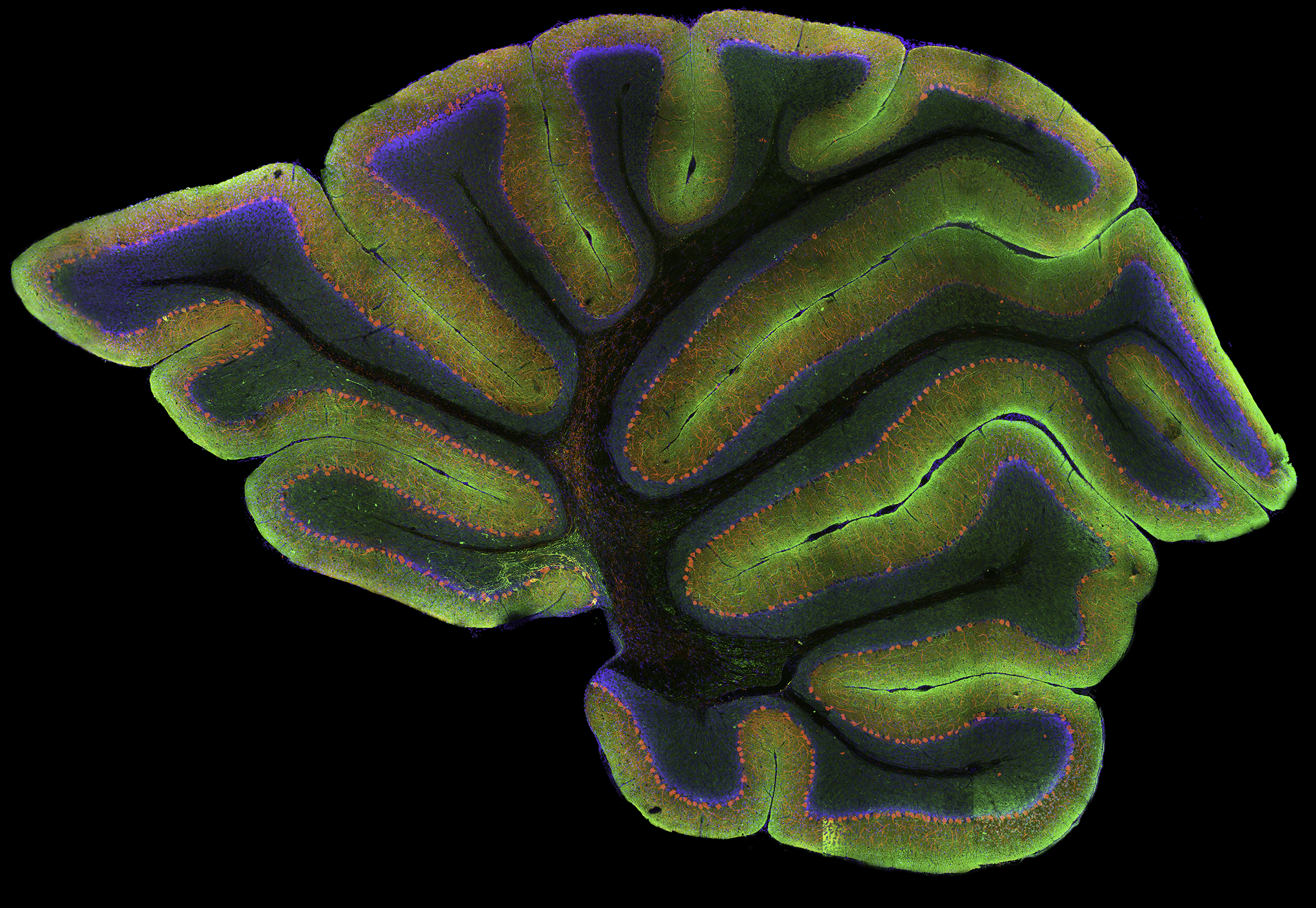
 High-resolution, wide-field mosaic of the cerebellum.
Each year I look forward to the Nikon Small World International Photomicrography competition. This year I was fortunate enough to receive image of distinction honors. The Small World competition featured thousands of entries from 89 countries, showcasing some of the worlds best microscopists who share their talents in the premiere imaging contest. I entered the image above of the cerebellum as part of my research in Dr. Kenneth Kosik’s cellular and molecular neurobiology lab in the Neuroscience Research Institute at UC Santa Barbara that was captured using immunoctyochemisty and a laser scanning confocal microscope. The resultant image is comprised of hundreds of individual images or tiles that are seamlessly aligned and registered to provide both a global overview of the specimen as well as the resolution necessary to interrogate selected regions at the single-cell level. The mosaic shows the Purkinje cells (red) lining the cerebellum, as well as the ubiquitous protein, tau (green) and nuclei (blue). I have always been drawn to the cerebellum in part because of its sulci and gyri that demonstrate a remarkable sense of evolutionary complexity and aesthetic beauty.
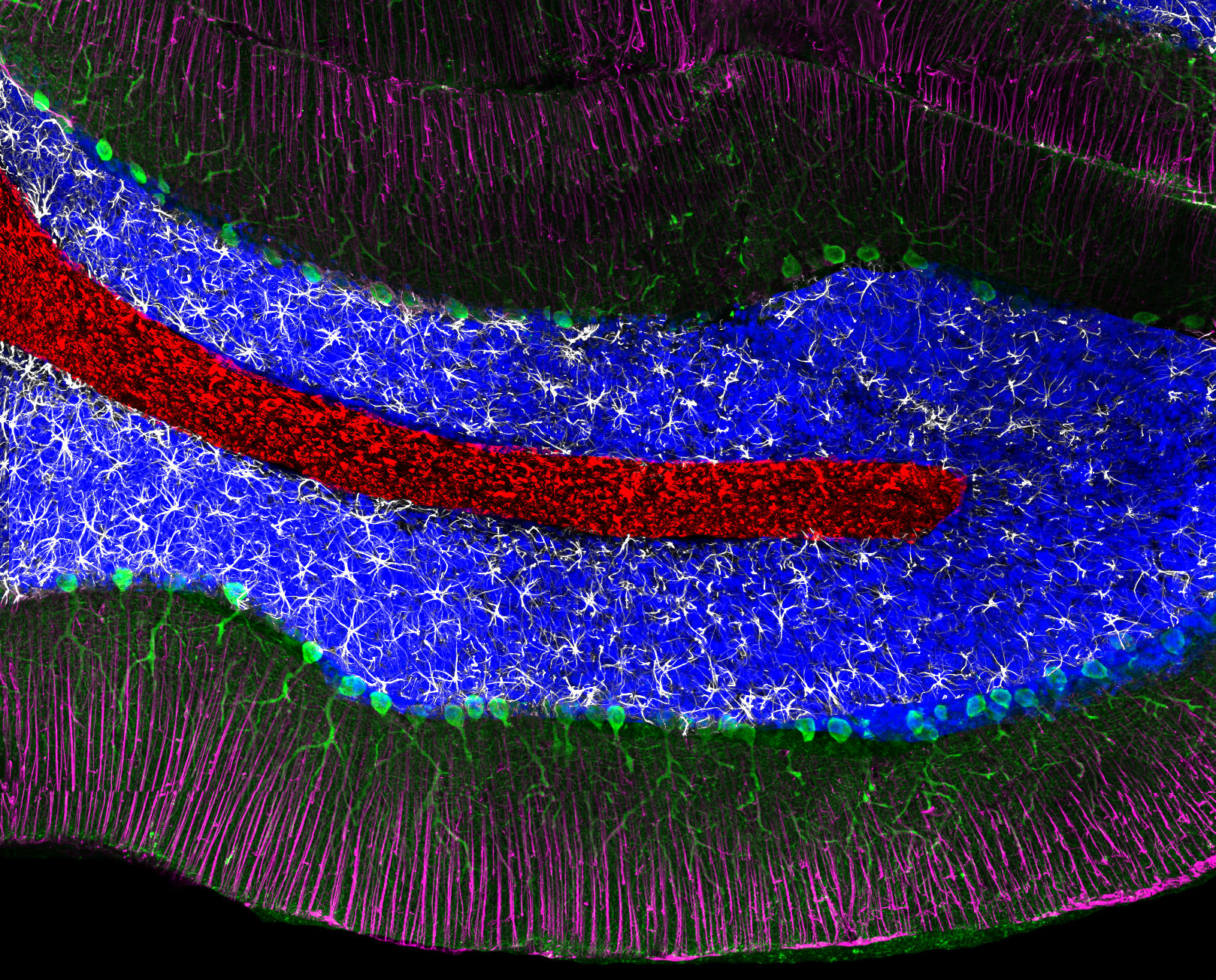 Example of a single tile that comprise the above mosaic.
This example shows the resolution of a single tile that comprises the winning image. Here, Perkinje cells stained with an antibody to inositol triphosphate 3 (green), the intermediate filament protein GFAP (glial fibrillary acidic protein; white), Bergmann’s glia (magenta), nuclei (blue) and myelin basic protein (red).