Cell-mediated remodeling of hydrogels triggered by adipogenic differentiation.

Adipose stem cells (immature fat cells) are among the most abundant cell-type in the human body, developing methodologies that take advantage of these cells for reconstructive procedures may one day provide a source for wound healing as a result of traumatic injury or to ameliorate congenital defects using autologous transplantation, thus obviating the potential of tissue rejection.  In a collaboration with Drs. Tracy Clevenger, Steve Fisher, Dennis Clegg we examined the ability of adipose stem cells to breakdown a hydrogel that was engineered to foster their growth and survival as these stem cells differentiated into a more mature state with the long-term rationale of increasing the chance of successful tissue grafting as well as accelerating the process of wound healing.  That research was published today in the Journal of Tissue Engineering.  This project involved a blend of computer science, molecular and cellular biology, as well as bio-imaging techniques to investigate these complex biological systems.

b-vnrgd-exp14-24hr-undiffmmp-slide-13_ipd-h244-1_mosaic

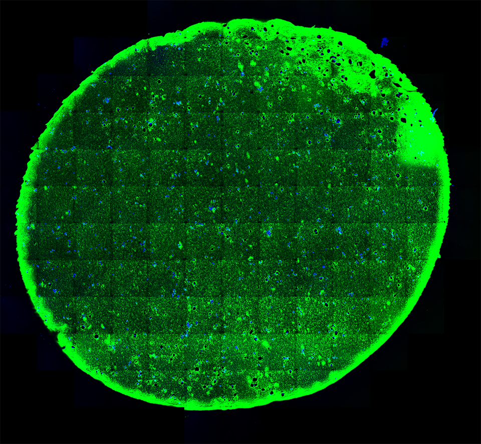
A 20-micron thick slice of a hydrogel immunostained (green; for reference the width of a human hair is about 180-microns) with embedded stem cells (blue) under “normal” conditions visualized using fluorescent microscopy.

ipd-14-slide-42-undiff-h_e-stain-20x-3

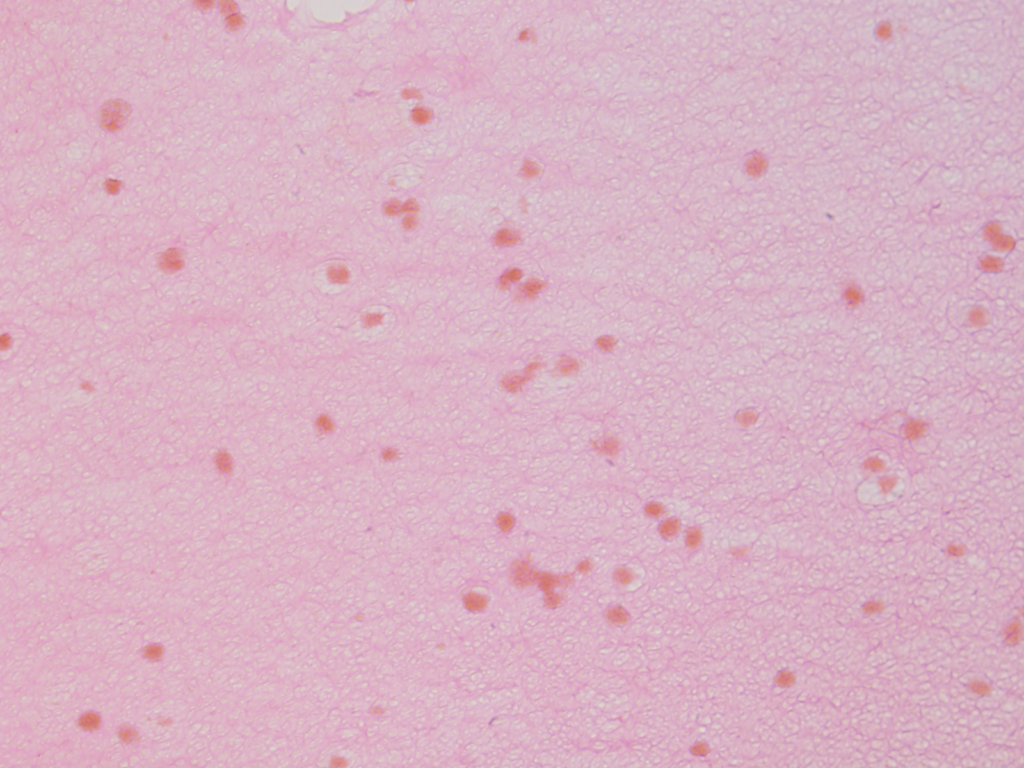
Using a stain that labels eosinophilic structures we are able to visualize the fine processes of hydrogel that uniformly interweave among the adipose stem cells using a light microscope.

b-vnrgd-exp13-4wk-diff-slide-32_ipd-h15-3_mosaic

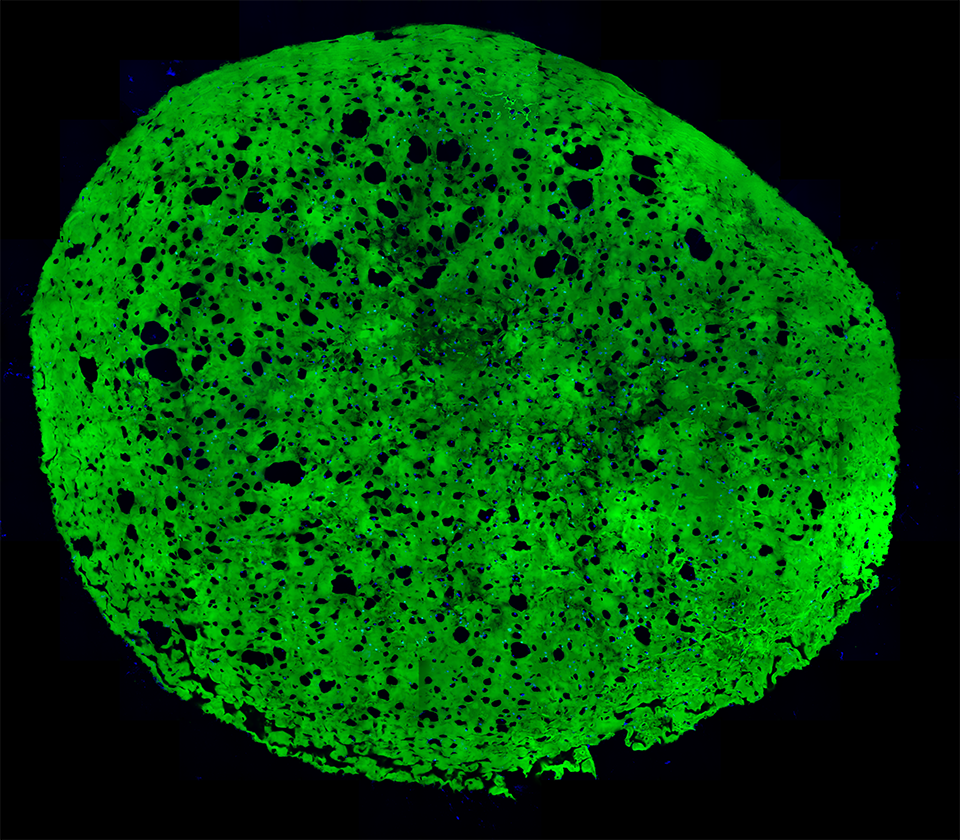
A section of a hydrogel whose embedded stem cells were placed under conditions that induced them to differentiate into more mature state (blue).  Notice the increase in the number of cavities after 4 weeks as a result of the adipogenic differentiation.

ipd-16-slide-70-diff-h_e-stain-20x-1

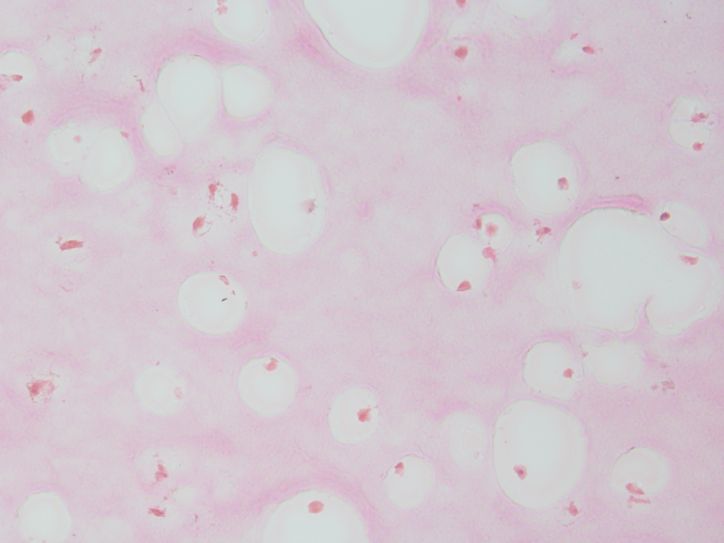
We can also documented the changes in the filamentous appearance of the hydrogel to a increasingly smoother appearance under differentiated conditions, shown in pink.

ipd-13-4wk-diff-mmp-slide-77-hoescht-b-map2-r-b-vnrgd-g-40x-p2-sb-20um

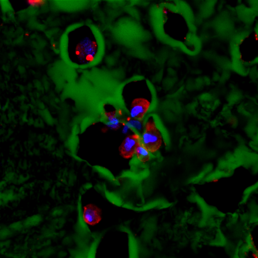
A high-resolution immunofluorescent image of a group of adipose stem cells lining the edges of a subset of cavities within the hydrogel.  Here, the gel is visible in green, while the cell nucleus is blue, and the cytoplasm of the cell is red.

b-vnrgd-exp11-4wk-diff-slide-30_ipd-h11-3_mosaic_cellcounting

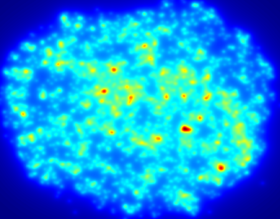
An example of a density map shows the relative distribution of cells across a section of hydrogel, here red areas show more densely populated regions. Data such as this illustrates the importance of undergraduate volunteers who contribute countless hours quantifying various parameters, those efforts help move research along at a faster pace than would otherwise happen.

web-prepared

Dr. Tracy Clevenger, lead author.

Cellular Remodeling and Genetic Changes Following Retinal Detachment

Fig 2 new

Fig 6 new

fig-8-new-for-nri-monitors

Fig 3 new

_MG_1605 FINAL_resized

Steve Fisher (Senior Author and Principal Investigator; Retinal Cell Biology Laboratory)

We have a new publication in a collaboration with Drs. Qirui Hu, Sheldon Miller, Peter Munson, Arvydas Maminiskis, and Bo Chang that examined the anatomical and genetic changes in the retina and underlying pigmented epithelium in a model of retinal detachment.  Retinal detachment initiates a cascade of changes at the cellular and genetic level, understanding these changes allow for the development of therapeutic agents aimed at arresting the neurodegenerative process.  That article in its entirety can be found here.

Abstract:
PURPOSE:  The purpose of this study was to examine the rpea1 mouse whose retina spontaneously detaches from the underlying RPE as a potential model for studying the cellular effects of serous retinal detachment (SRD).
METHODS:  Optical coherence tomography (OCT) was performed immediately prior to euthanasia; retinal tissue was subsequently prepared for Western blotting, microarray analysis, immunocytochemistry, and light and electron microscopy (LM, EM).
RESULTS:  By postnatal day (P) 30, OCT, LM, and EM revealed the presence of small shallow detachments that increased in number and size over time. By P60 in regions of detachment, there was a dramatic loss of PNA binding around cones in the interphotoreceptor matrix and a concomitant increase in labeling of the outer nuclear layer and rod synaptic terminals. Retinal pigment epithelium wholemounts revealed a patchy loss in immunolabeling for both ezrin and aquaporin 1. Anti-ezrin labeling was lost from small regions of the RPE apical surface underlying detachments at P30. Labeling for tight-junction proteins provided a regular array of profiles outlining the periphery of RPE cells in wild-type tissue, however, this pattern was disrupted in the mutant as early as P30. Microarray analysis revealed a broad range of changes in genes involved in metabolism, signaling, cell polarity, and tight-junction organization.
CONCLUSIONS:  These data indicate changes in this mutant mouse that may provide clues to the underlying mechanisms of SRD in humans. Importantly, these changes include the production of multiple spontaneous detachments without the presence of a retinal tear or significant degeneration of outer segments, changes in the expression of proteins involved in adhesion and fluid transport, and a disrupted organization of RPE tight junctions that may contribute to the formation of focal detachments.