Bioengineered scaffolds and adipose-derived stem cells

4wk Diff -MMP B-VnRGD (r) Map2 (g) Hoescht (b) p3_PS

B-VnRGD Exp#13 4wk Undiff +MMP slide 10_IPD-H14.3_mosaic

IPD 6 4wk UnDiff -MMP slide 25 Hoescht (b) MAP2 (r) B-VnRGD (g) 20x p2

Animal Exp#4 InVitro #3 12wk Diff ++slide 79.2_mosaic

IPD 13 4wk Diff +MMP slide 5 Hoescht (b) MAP2 (r) B-VnRGD (g) 20x p1_PS

Web Prepared

10348883_10104465063079627_8804975298544853794_o

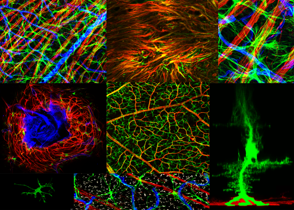
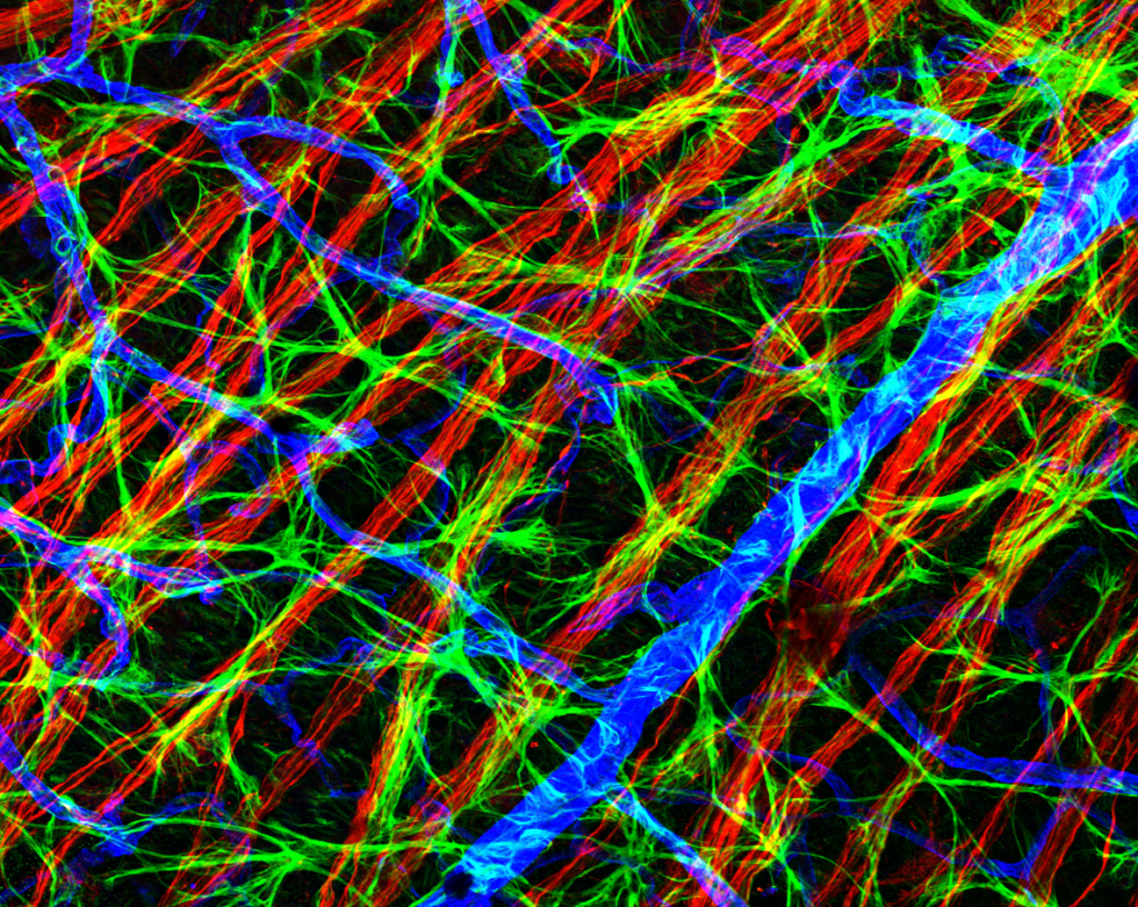
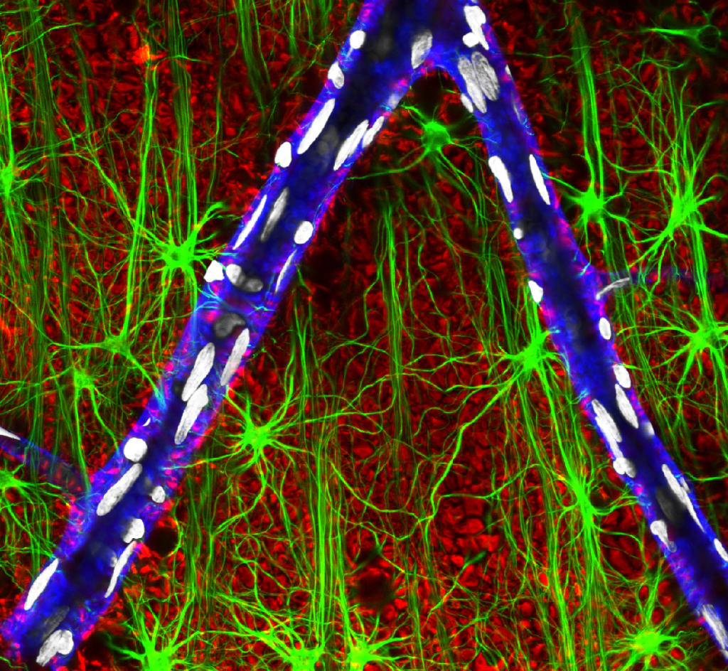
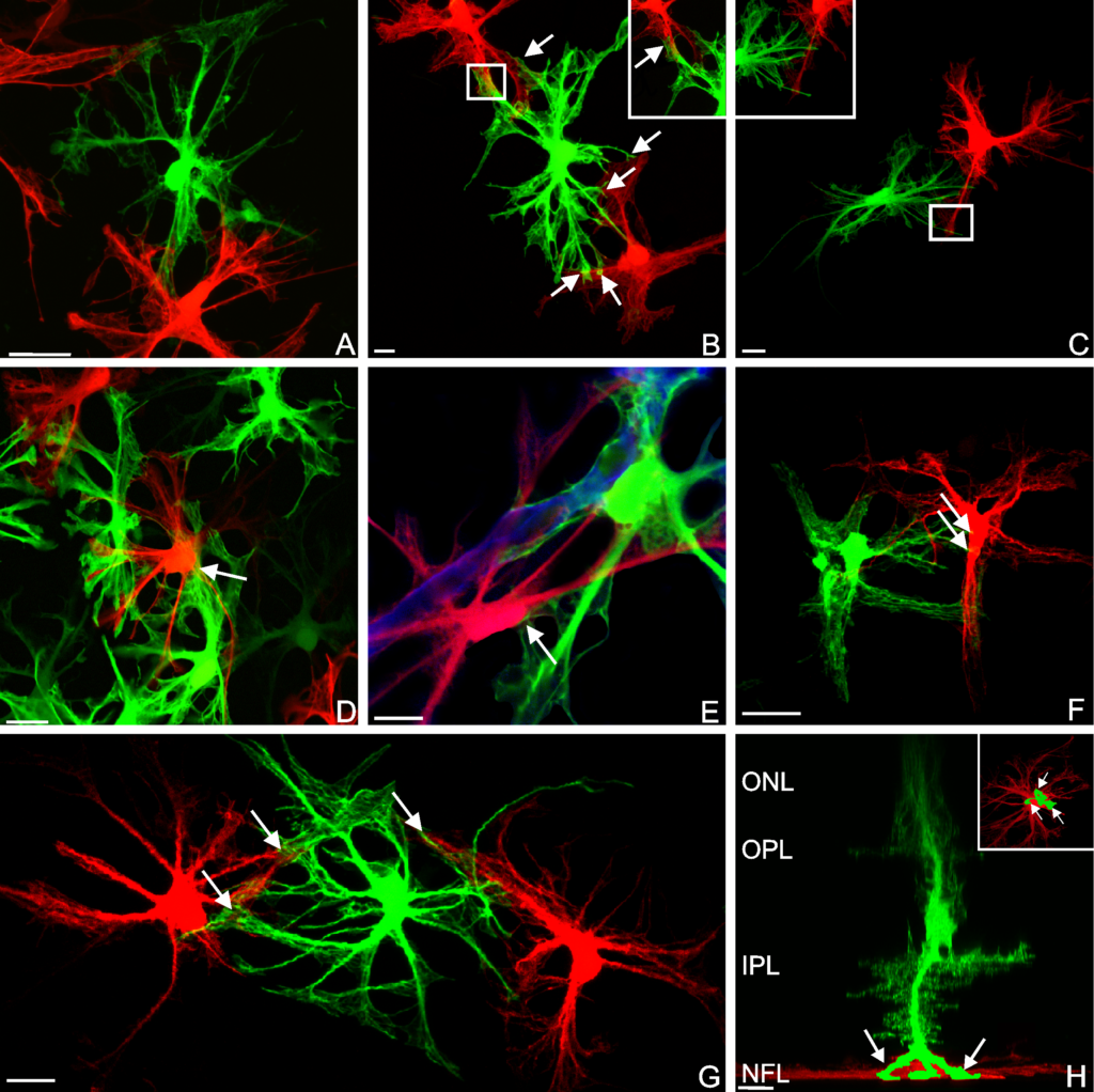
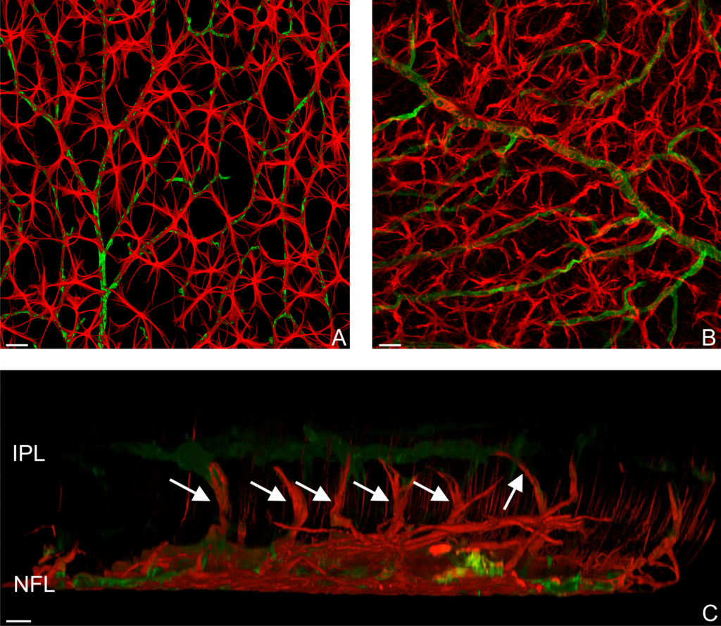
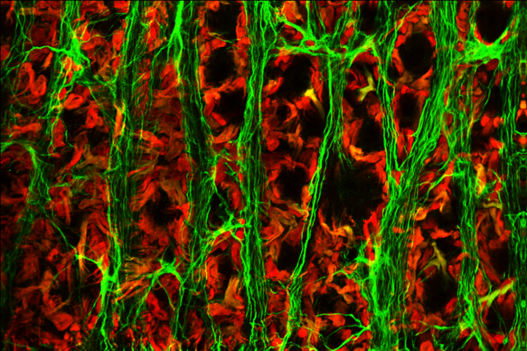
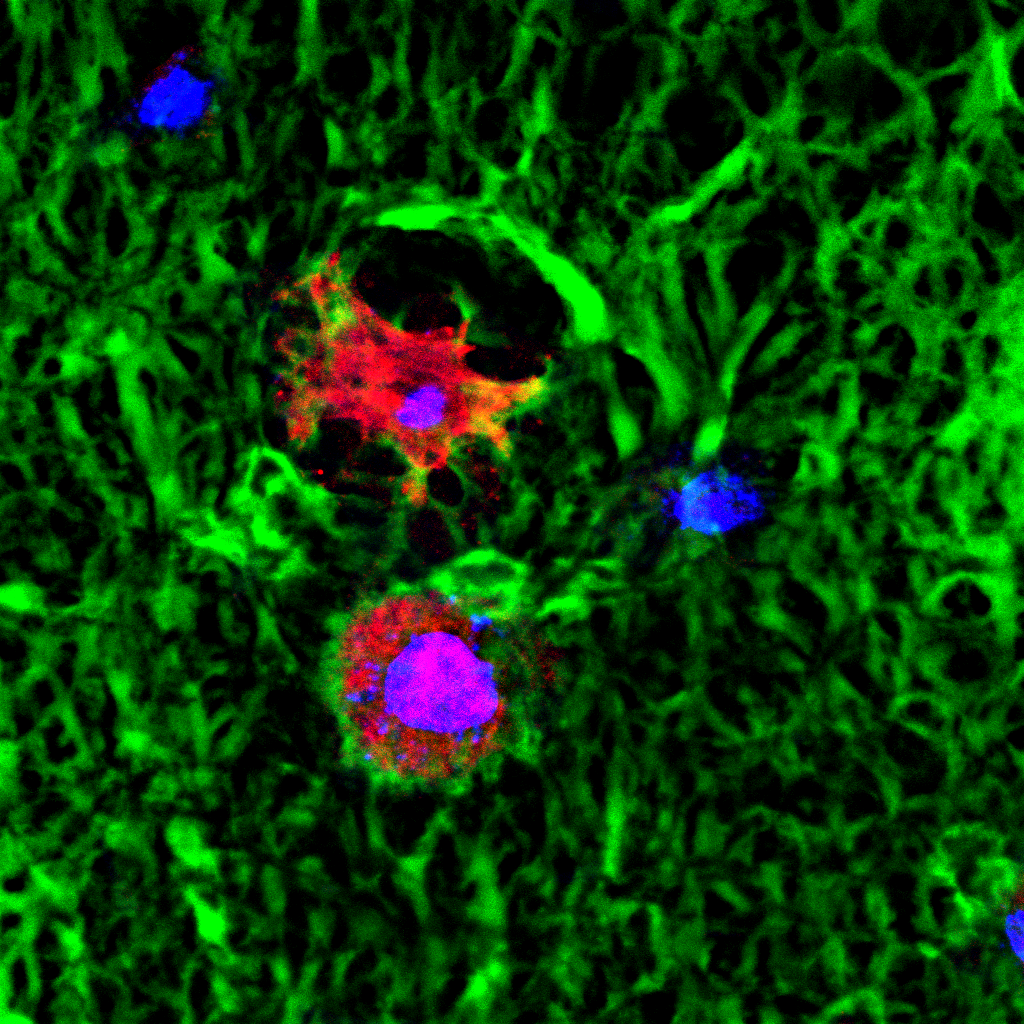
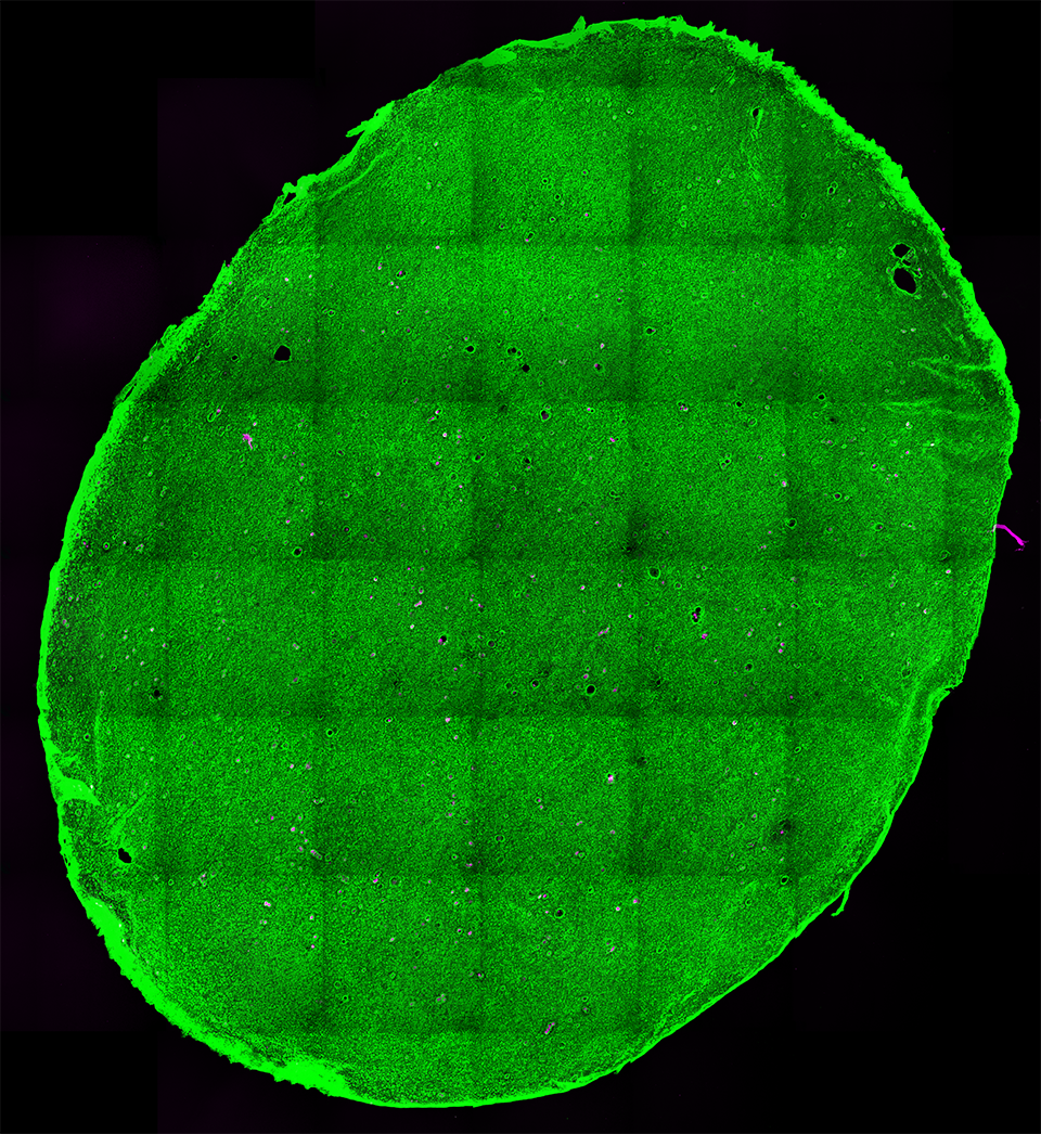
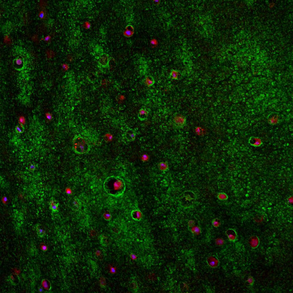
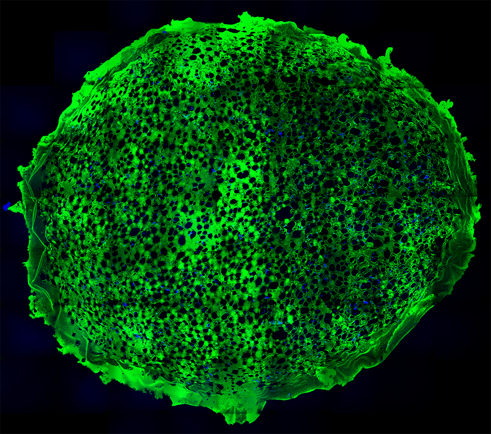
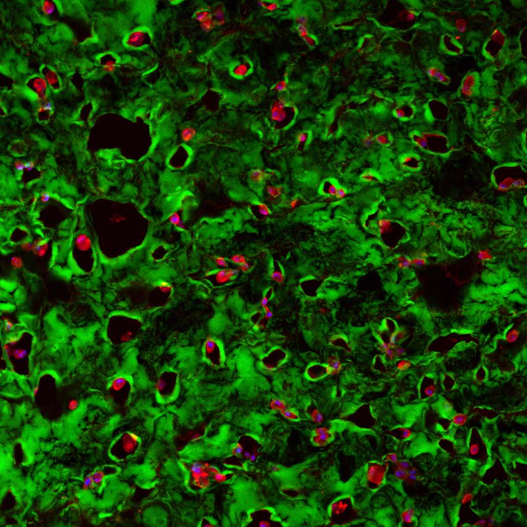
     New publication! Drs. Tracy Clevenger, Steve Fisher, Dennis Clegg and myself recently reviewed wide-ranging strategies concerning synthetic scaffolds and their potential to foster adipose-derived stem cells as therapies involving soft-tissue, bone, and cartilage, that work can be found here.  Research involving adipose-derived stem cells has potential implications in a wide-range of applications encompassing cosmetic procedures, burn patients, and wound healing, however, research involving these cells remains in its infancy.  Microscopy images above show examples of these synthetic systems harboring stem cells, in these images the green color corresponds to the synthetic scaffold, the blue indicates the nucleus of an individual adipose-derived stem cell, lastly the red displays a part of the cells’ cytoskeleton that in part reveals its “shape”.


     Abstract:  Regenerative medicine possesses the potential to ameliorate damage to tissue that results from a vast range of conditions, including traumatic injury, tumor resection and inherited tissue defects. Adult stem cells, while more limited in their potential than pluripotent stem cells, are still capable of differentiating into numerous lineages and provide feasible allogeneic and autologous treatment options for many conditions. Adipose stem cells are one of the most abundant types of stem cell in the adult human. Here, we review recent advances in the development of synthetic scaffolding systems used in concert with adipose stem cells and assess their potential use for clinical applications.

Astrocyte Reactivity and Plasticity

Post 1

Lasker 4 month RE_mosaic2

cover image EER

Fig 3

Fig 10

NWGVP2 copy

Recently, Drs. Patrick Keeley, Ben Reese, Geoff Lewis, Steve Fisher, and myself published a review article in the Journal of Experimental Eye Research examining the reactive nature of retinal astrocytes in response to injury.  The imagery above comes from that publication, which can be found on PubMed here. A synopsis of the issue and the article itself, as well as the other articles accompanying this special issue on retinal remodeling can also be found here.

Abstract:

Although retinal neurodegenerative conditions such as age-related macular degeneration, glaucoma, diabetic retinopathy, retinitis pigmentosa, and retinal detachment have different etiologies and pathological characteristics, they also have many responses in common at the cellular level, including neural and glial remodeling. Structural changes in Müller cells, the large radial glia of the retina in retinal disease and injury have been well described, that of the retinal astrocytes remains less so. Using modern imaging technology to describe the structural remodeling of retinal astrocytes after retinal detachment is the focus of this paper. We present both a review of critical literature as well as novel work focusing on the responses of astrocytes following rhegmatogenous and serous retinal detachment. The mouse presents a convenient model system in which to study astrocyte reactivity since the Mϋller cell response is muted in comparison to other species thereby allowing better visualization of the astrocytes. We also show data from rat, cat, squirrel, and human retina demonstrating similarities and differences across species. Our data from immunolabeling and dye-filling experiments demonstrate previously undescribed morphological characteristics of normal astrocytes and changes induced by detachment. Astrocytes not only upregulate GFAP, but structurally remodel, becoming increasingly irregular in appearance, and often penetrating deep into neural retina. Understanding these responses, their consequences, and what drives them may prove to be an important component in improving visual outcome in a variety of therapeutic situations. Our data further supports the concept that astrocytes are important players in the retina’s overall response to injury and disease.