We have a new publication out in the journal of Investigative Ophthalmology and Visual Science that examined cellular and sub-cellular remodeling in the hibernating ground squirrel. This research was a result of another fruitful collaboration with Ben Sajdak, a graduate student in the department of advanced ocular imaging program at the medical college of Wisconsin Eye Institute. Open access PDF can be found here.
Authors: Benjamin S. Sajdak, Brent A. Bell, Taylor R. Lewis, myself, Grayson S. Cornwell, Steven K. Fisher, Dana K. Merriman, and Joesph Carroll.
Abstract:
Purpose. We examined outer retinal remodeling of the euthermic and torpid cone-dominant 13-lined ground squirrel (13-LGS) retina using optical coherence tomography (OCT) imaging and histology.
Methods. Retinas and corneas of living 13-LGSs were imaged during euthermic and torpid physiological states using OCT. Retinal layer thickness was measured at the visual streak from registered and averaged vertical B-scans. Following OCT, some retinas were collected immediately for postmortem histologic comparison using light microscopy, immunofluorescence, or transmission electron microscopy.
Results. Compared to OCT images from euthermic retinae, OCT images of torpid retinae revealed significantly thicker inner and outer nuclear layers, as well as increases in the distances between outer retinal reflectivity bands 1 and 2, and bands 3 and 4. A significant decrease in the distance between bands 2 and 3 also was seen, alongside significant thinning of the choriocapillaris and choroid. OCT image quality was reduced in torpid eyes, partly due to significant thickening of the corneal stroma during this state.
Conclusions. The torpid retina of the hibernating 13-LGS undergoes structural changes that can be detected by OCT imaging. Comparisons between in vivo OCT and ex vivo histomorphometry may offer insight to the origin of hyperreflective OCT bands within the outer retina of the cone-dominant 13-LGS.
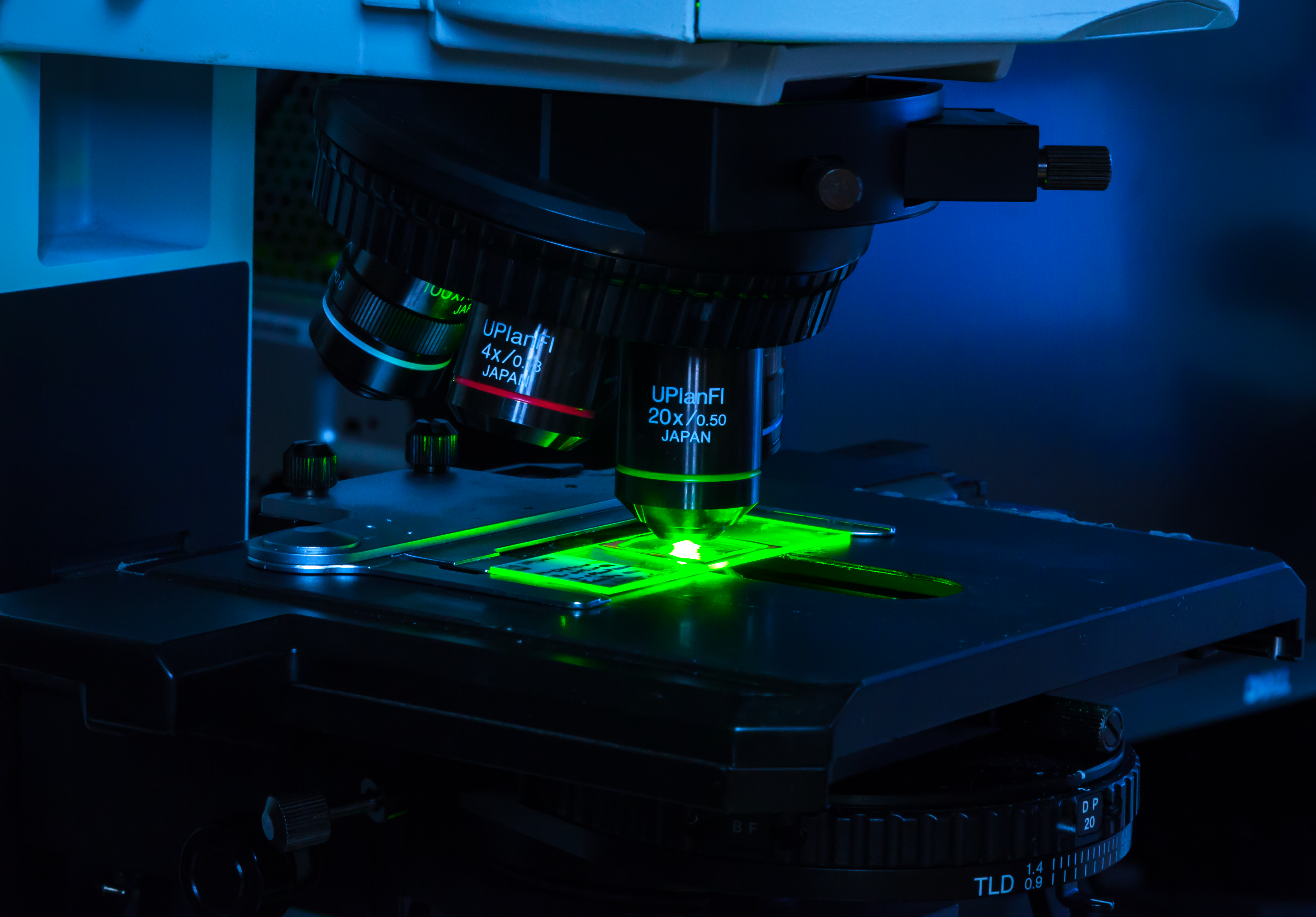 Olympus flouview 1000 laser scanning confocal microscope.
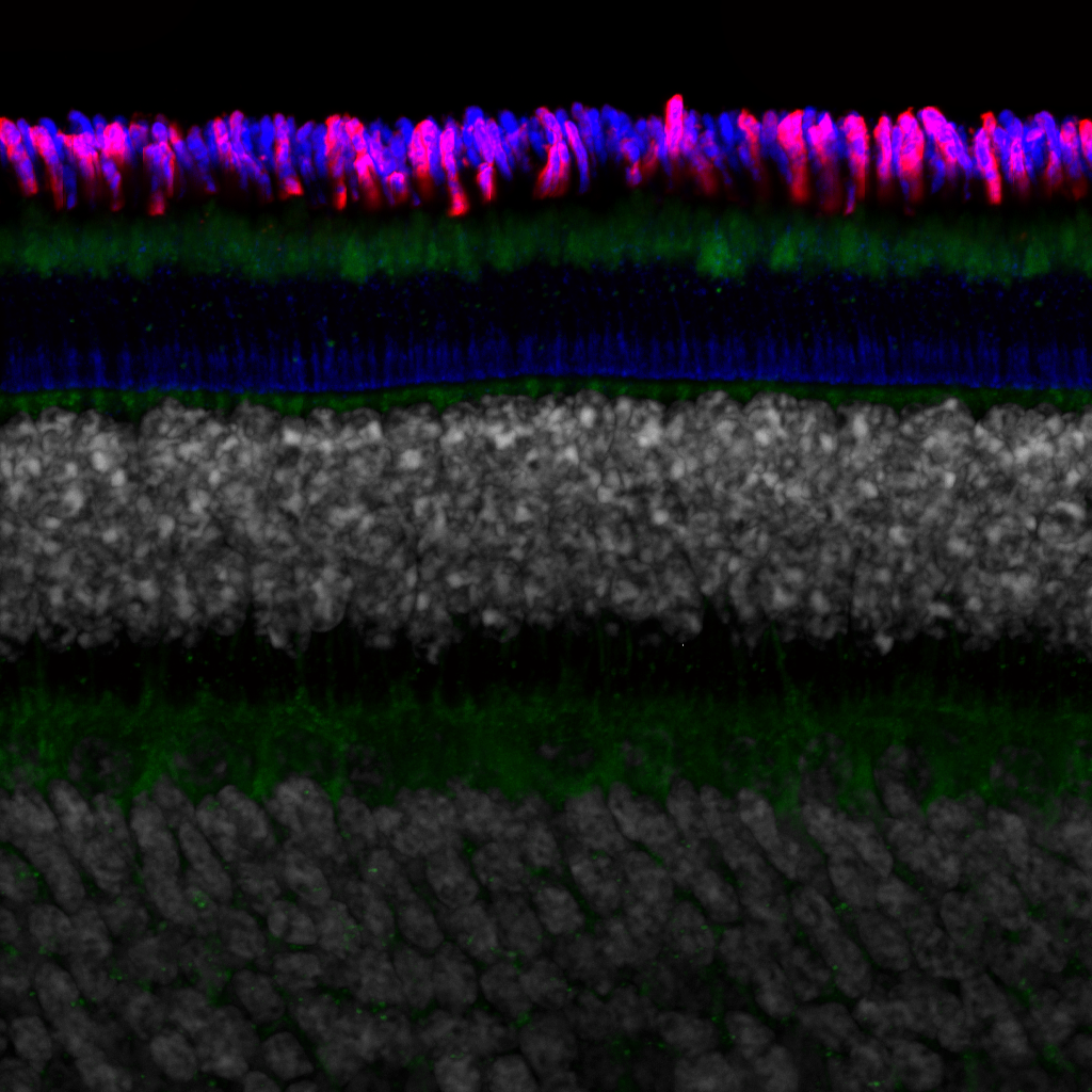
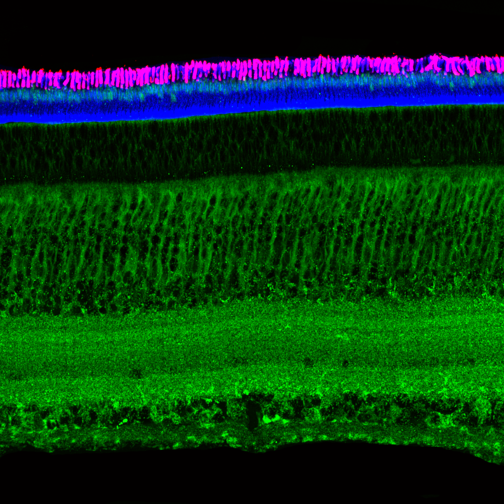 Light micrograph using immunocytochemistry to visualize the cone photoreceptors of the neural retina, magnified 400 times.
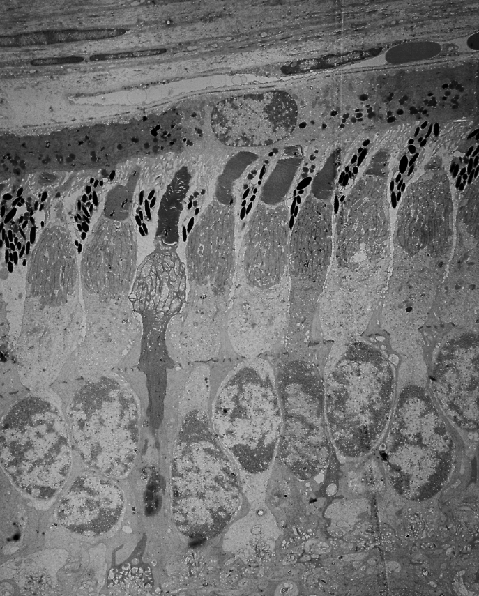 Electron micrograph of the light sensitive photoreceptors, magnified 10,000 times.
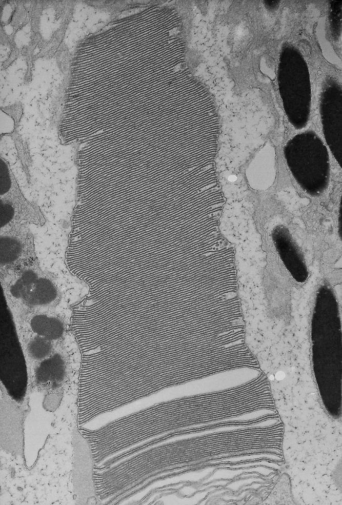 Electron micrograph of a photoreceptor outer segment, magnified 20,000 times.